聚焦意识变化、瞳孔、颈椎保护、气道风险和团队报告语言。页面只训练观察与复评,本页提供有来源的参考数字与关键步骤:实习识别与报告;规培在上级监督下执行;主治指挥与资源调度。剂量与阈值;以本院规范为准。Focus on mental-status change, pupils, c-spine protection, airway risk, and team reporting language. The page trains observation and reassessment only, not intubation thresholds or medication plans.
source_required
预计学习Estimated learning time · 30 分钟min
学完这门课,你能……After this course, you can…
报告意识、瞳孔及 GCS 分项变化。Report consciousness, pupils and GCS component changes.
将神经变化与氧合、通气、灌注一起解释。Interpret neurological change with oxygenation, ventilation and perfusion.
区分来源影像、合成病例与患者个体决策。Distinguish source imaging, simulation and patient-specific decisions.
实习:识别与报告;规培:决策时机与监督下关键步骤;青年主治:团队指挥与资源调度。Intern: recognize/report; resident: decision timing and supervised key steps; early attending: team leadership and resources.
颅脑损伤:从意识变化开始TBI: begin with changing consciousness
意识在下降,先救哪一条生理线?Consciousness is declining: which physiologic threat comes first?
沿用本课合成病例,非真实病历。逐条释放是学习节奏;临床立即威胁并行处理。选择不会改写患者生理。Retained synthetic teaching case, not a patient record. Release structures learning; immediate threats require parallel care. Choices never rewrite physiology.
主诉Presenting complaint
头部撞击后短暂意识丧失,伴反应与语言改变。Brief loss of consciousness after head impact, with altered response and speech.
现病史History
电动车碰撞后头部着地;伤前神经状态、确切伤后时间、抗凝用药及过敏史待核实。Head impact after an e-bike collision; baseline neurology, exact injury time, anticoagulants and allergies need confirmation.
院前交接Prehospital handoff
颈椎保护、氧疗及用药情况原病例未详记;交接时逐项询问,不能假定已完成。Spinal precautions, oxygen and drugs were not fully documented; verify each during handoff rather than assume completion.
先核对初步查体与未知项Initial examination and unknowns
GCS 14(E4V4M6),双瞳孔 3 mm 等大灵敏。GCS 14 (E4V4M6); pupils 3 mm, equal and reactive.
未提供的阴性体征不是已经排除;补问用药、氧疗及观察条件。An unreported negative finding is not an exclusion; verify medication, oxygen and observation context.
首组氧疗条件原记录未明确;体温为合成补充。Initial oxygen context was not recorded; temperature is a synthetic addition.
※为本轮合成教学补充,并非从真实病历追回的数据。氧疗条件是病例设定,不是推荐剂量;数值随预设观察时点变化,不由答题决定。※ marks synthetic teaching additions, not recovered patient records. Oxygen context is a case assumption, not a prescribed dose; preset observations change by time, never by quiz answer.
血糖/检验学习单:区分原值与教学补充Laboratory learning sheet: originals versus additions
到院0分钟教学补充,非原始病历结果;这些正常示例不排除颅内进展。血气、凝血与用药背景仍需按临床获得。Arrival teaching additions, not original records. Normal examples do not exclude intracranial progression; obtain gas, coagulation and drug context as indicated.
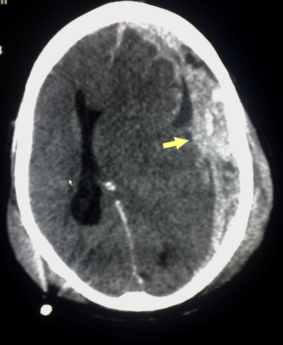
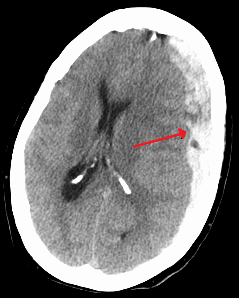
两位外部患者、两个观察任务:找血肿,再找占位效应。不要把它们拼成恶化时序。Two external patients, two tasks: identify haemorrhage, then mass effect. Do not combine them into a deterioration timeline.
外部病例 A:创伤性硬膜下出血 CTExternal case A: traumatic subdural haemorrhage CT
先看哪里Where to look
寻找靠近颅骨内板的高密度新月形区域;本图异常在显示右侧,即患者左侧。先确认方位,再描述形态。Look for a hyperdense crescent along the inner skull. The abnormality is on image right, patient left. Confirm orientation before describing morphology.
这张图不能回答What this image cannot answer
静态切片不能确定 GCS、ICP、当前是否脑疝或治疗指征。它不是下图患者的早期检查。A single slice cannot determine GCS, ICP, current herniation or treatment indications. It is not an earlier scan of the next patient.
原始图注与许可Original caption and licence
Glitzy queen00(英文维基 English Wikipedia);箭头标注由 Delldot 后期添加 · Public domain(公有领域) · Original source / 原图出处
This CT scan is an example of Subdural haemorrhage caused by trauma.
保留原图;网页仅按比例显示。读图释义由课程另写,不是原作者诊疗结论。Original image retained and displayed proportionally. Teaching interpretation is separate from the original clinical conclusions.
外部病例 B:硬膜下血肿伴中线移位External case B: subdural haematoma with midline shift
先看哪里Where to look
比较左右半球、脑室形态与中线;红箭指向患者左侧高密度血肿。Compare hemispheres, ventricular shape and midline; the red arrow indicates a left-sided hyperdense collection.
这张图不能回答What this image cannot answer
中线移位是占位效应线索,不能单凭图片决定手术。与上图是不同患者。Shift is a clue to mass effect, not a standalone surgical indication. This is a different patient from the preceding image.
保留原图;网页仅按比例显示。读图释义由课程另写,不是原作者诊疗结论。Original image retained and displayed proportionally. Teaching interpretation is separate from the original clinical conclusions.
避免低氧和低血压;颅内监测、重复影像及手术评估结合伤情、趋势与神经外科判断,不能由单张 CT 自动触发。Avoid hypoxaemia and hypotension. Monitoring, repeat imaging and surgical review depend on injury, trajectory and neurosurgical assessment, not one CT alone.
03
复评要可比较Make reassessment comparable
连续记录 GCS 的 E/V/M、瞳孔与四肢反应,同时记录全身生理。新变化比“已经做过 CT”更能提示重新评估。Record GCS components, pupils and limb responses with systemic physiology. New changes require reassessment even after CT.
暂停判断:GCS 下降,但 CT 刚完成,能等下一班吗?Pause and decide: GCS falls soon after CT. Can review wait for the next shift?
不能以检查已完成结束复评。核对混杂因素,同时升级神经评估并明确重复影像的临床触发点。A completed scan does not end reassessment. Check confounders while escalating neurologic assessment and specifying triggers for repeat imaging.
真实公开图用于观察训练;病例与指南各自标明来源,病例报告不能单独建立普遍治疗规则。Public images support observation training. Case reports and guidelines are distinguished; a case report alone cannot establish a general treatment rule. Clinical applicability remains pending teacher review.
BEDSIDE → SPECIALIST → REASSESS
床旁救治与专科接续Bedside care and specialist continuity
每项措施都要回答:为什么做、由谁做、做后看什么。侵入操作由受训获授权人员执行,学员在监督下训练。For every action: why, who and what to reassess. Trained authorised operators perform invasive care; learners practise under supervision.
01
床旁:保护脑与气道Bedside: protect brain and airway
先纠正低氧和低灌注,维持颈椎保护;有分泌物处理困难、通气或气道保护不足时由高级团队评估气道。Correct oxygenation and perfusion while protecting the cervical spine; advanced teams assess airway protection and ventilation when inadequate.
02
恶化:复查与专科Decline: re-examine and escalate
新意识、瞳孔或运动变化立即复评并讨论再影像;CT阴性不能替代持续观察。New mental, pupil or motor changes require urgent reassessment and consideration of repeat imaging; a negative CT does not replace observation.
占位病变、颅压监测、分层颅压治疗与手术由专科结合临床和CT决定;不把常规过度通气作为普遍措施。Specialists integrate examination and CT for mass lesions, ICP monitoring, tiered therapy and surgery; routine hyperventilation is not a universal measure.
步骤、适用条件与复评核查Sequence, conditions and reassessment checks
确认主导威胁与当前生理,口头分派操作者和监护者。Confirm the dominant threat and current physiology; name operator and monitor.
按院内规程准备器材、通路、血液及专科资源;救命处置不等教学问答。Prepare equipment, access, blood and specialist resources under local protocol; rescue care never waits for teaching prompts.
由获授权团队实施所选措施;必要时结合技能页的专业步骤,不能用流程卡代替操作培训。Authorised teams deliver the selected intervention; linked skills supply procedure teaching, not certification by a flow card.
立即比较操作前后氧合、灌注与意识,记录时间、氧疗、药物及失败信号。Immediately compare oxygenation, perfusion and consciousness and record time, oxygen, drugs and failure signals.
没有改善时重新寻找未解决机制,升级专科或替代路径,明确下一复评责任人。If response is inadequate, reassess mechanisms and escalate specialists or fallback pathways with a named next owner.
来源、版本与教学解释Sources, versions and teaching interpretation
2026-10-04核对公开原始资料;标注文件实际年份,不统称“2026最新”。本页为教学整理,不复制官方算法;新增内容待临床教师终审。Public primary sources checked 2026-10-04; document years are shown rather than labelled “latest 2026”. This is teaching synthesis, not a copied official algorithm. New content awaits clinical teacher review.
A/D 联动总览图A/D Linkage Overview
把颅脑损伤学习从“诊断名”转成气道、颈椎保护、神经变化和复评路线。Turn TBI learning from a diagnosis label into airway, c-spine, neurologic change, and reassessment route.
visual_asset_pending_review
01 · OBSERVE意识、语言与瞳孔Mental status, voice, pupils把当前可见事实与此前状态分开描述。Separate current facts from the prior state.
02 · LINK A/DA 与 D 同时进入表征Link A and D神经变化是否同时改变气道风险语言。Ask whether neurologic change alters airway-risk language.
03 · CONTEXT机制与颈椎信息Mechanism and c-spine补入保护状态、症状和仍未知的信息。Add protection status, symptoms, and unknowns.
04 · REASSESS检查后重写风险Rewrite risk after new information影像或会诊信息不能替代床边趋势。Imaging or consultation cannot replace bedside trend.
神经状态趋势板Neurologic trend board
MENTAL↓与前次比较versus prior
PUPILSΔ新变化new change
VOICEΔ报告事实report fact
RESPONSE↓触发复评reassess
01描述事实Describe facts
02对比变化Compare changes
03明确复评Plan reassessment
观察线索Observation cues
情境图用于看“变化从哪里被发现”,趋势板用于看“变化是否持续”。The scene shows where change is noticed; the trend board shows whether it persists.
source_required
01描述事实Describe facts
02对比变化Compare changes
03明确复评Plan reassessment
复评问题Reassessment question
新信息是否同时改变 A/D 风险、颈椎保护语言和下一信息需求。Whether new information changes A/D risk, c-spine language, and the next information need.
把机制、连续神经查体、全身生理、影像变化和团队升级放进同一时间轴。Place mechanism, serial neurologic examination, systemic physiology, imaging evolution, and escalation on one timeline.
ACS TBI 2024ACS TBI 2024
教材式核心Textbook core原发性损伤发生在受力当时,随后低氧、低灌注、通气异常、颅内病变进展、发热和代谢紊乱可加重继发性损伤。学习重点不是背一次 GCS,而是描述每个分项、瞳孔、局灶体征、语言/气道保护能力及其连续变化。Primary injury occurs at impact; subsequent hypoxemia, hypoperfusion, ventilatory disturbance, expanding intracranial lesions, fever, and metabolic derangement can amplify secondary injury. The goal is not one memorized GCS total, but serial description of components, pupils, focal signs, voice/airway protection, and change.
连续神经查体Serial neurologic examination
分别记录睁眼、语言和运动反应,比较双侧瞳孔大小与反应、肢体差异、抽搐、躁动/嗜睡和气道分泌物处理能力;并行核查镇静、低氧、低灌注和低血糖等混杂因素,同时评估颅内恶化。Record eye, verbal, and motor responses separately; compare pupils, limb asymmetry, seizure, agitation/drowsiness, and secretion handling. First consider confounders such as sedation, hypoxemia, hypoperfusion, and hypoglycemia.
影像与趋势Imaging and trend
头颅 CT 用于识别出血、占位效应、骨折和其他急性改变;一次阴性 CT 不能否定全部 TBI,也不能替代神经状态持续恶化时的重新评估。Head CT identifies hemorrhage, mass effect, fracture, and other acute changes. A negative CT does not exclude all TBI and cannot replace renewed evaluation when neurologic status worsens.
团队处置框架Team management frame
同步维持氧合、通气和灌注,保护颈椎,及早识别可逆全身原因;神经恶化、瞳孔改变或影像高风险信息应推动高级监护、神经外科和重复影像等团队升级。Maintain oxygenation, ventilation, and perfusion while protecting the cervical spine and correcting reversible systemic causes. Neurologic decline, pupil change, or high-risk imaging should prompt team escalation to advanced monitoring, neurosurgical review, and repeat imaging as appropriate.
复评句式Reassessment phrase
“与上次相比,E/V/M、瞳孔、局灶体征、气道保护、氧合和循环分别怎样变化?”“Compared with last time, how have E/V/M, pupils, focal signs, airway protection, oxygenation, and circulation changed?”
常见陷阱Common trap
把躁动只归因于“不配合”,或看到一次正常影像后停止观察;两者都会漏掉动态恶化。Attributing agitation to noncooperation or stopping observation after one reassuring image can both miss evolving deterioration.
交接重点Handover focus
报告基线、最近一次变化、时间点、已排除的混杂因素、当前保护状态以及下一次复评责任人。Report baseline, latest change and time, addressed confounders, current protection status, and ownership of the next reassessment.
记录意识、语言、瞳孔、运动反应及其与前次的差异。Record mental status, voice, pupils, motor response, and difference from prior.
产出:当前事实 + 前后变化Output: current facts + change
02报告Report
用短句同时表达 A/D 风险和颈椎背景,不使用未经确认的诊断标签。Report A/D risk and c-spine context without an unconfirmed label.
产出:一句趋势报告Output: one trend report
03整合资源Integrate
说明影像、上级团队和转运信息分别回答什么、仍留下什么未知。State what imaging, senior support, and transfer information answer and leave unresolved.
产出:信息节点清单Output: information-node list
04复评Reassess
比较新旧神经状态并重新明确主导风险和下一次复评责任。Compare serial neurologic status and restate dominant risk and reassessment ownership.
产出:下一次复评问题Output: next reassessment question
来源与审核状态Sources and Review Status
连续神经查体、全身生理与继发性损伤、影像边界和团队升级框架已按 2024 ACS TBI 最佳实践核对;具体监护与治疗流程仍需院内专家审核。Serial neurologic examination, systemic physiology and secondary injury, imaging limits, and escalation were checked against the 2024 ACS TBI best-practice guideline; institutional monitoring and treatment pathways still require expert review.
CRASH-3招募GCS≤12或CT证实颅内出血、无严重颅外出血;轻中度亚组结果不能推广为所有TBI获益。CRASH-3 enrolled GCS at most12 or CT-confirmed intracranial bleeding without major extracranial hemorrhage; subgroup benefit is not universal TBI benefit.
防继发损伤:年龄、氧合与灌注Prevent secondary injury: age, oxygenation and perfusion
先报告E/V/M与瞳孔Report E/V/M and pupils
重型TBI:SBP≥100(50–69岁),≥110(15–49岁或>70岁);国内MAP≥80 mmHg。CPP60–70 mmHg、ICP>22 mmHg需处理;BTF压力证据III级,ICP/CPP为IIB级。以本院规范为准。Severe TBI: SBP at least 100 at 50–69 years, at least 110 at 15–49 or over 70; Chinese MAP at least 80 mmHg. CPP60–70, treat ICP over 22; BTF pressure evidence LevelIII, ICP/CPP LevelIIB. Follow institutional policy.
GCS≤8、保护反射丧失或脑疝提示气管插管评估;同时预氧合、SMR、纠正低血压。报GCS分项、不可评估因素、双瞳大小/反射;GCS-P=总分减无反应瞳孔数。以本院规范为准。GCS at most8, lost protective reflexes or herniation prompts airway/intubation evaluation with preoxygenation, SMR and pressure correction. Report components, confounders and both pupils; GCS-P subtracts the count of unreactive pupils. Follow institutional policy.
国内SpO₂至少90%、理想>95%;ACS目标≥94%。无颅内高压时PaCO₂/PetCO₂参考35–45 mmHg,休克时EtCO₂不能替代动脉血气。以本院规范为准。Chinese SpO₂ floor90%, ideally over 95%; ACS target at least 94%. Without intracranial hypertension, PaCO₂/PetCO₂ reference35–45 mmHg; shock invalidates assuming EtCO₂ equals arterial CO₂. Follow institutional policy.
脑疝可短时救援通气,先35–38、必要时32–35 mmHg,严密监测并连接确定性治疗;不能对所有TBI持续降CO₂。以本院规范为准。Herniation permits brief rescue ventilation, initially35–38, if necessary32–35 mmHg, with close monitoring and definitive treatment; do not persistently lower CO₂ for all TBI. Follow institutional policy.
甘露醇采用ENLS成人0.5–1 g/kg参考;国内原文20%制剂mL/kg与括注g/kg存在换算不一致,不能照抄体积。按本院制剂、渗透压和血压核算;BTF第4版未确立优选药/剂量。以本院规范为准。Use ENLS adult mannitol0.5–1 g/kg reference. The Chinese text contains inconsistent20% mL/kg versus g/kg conversion: do not copy the volume. Verify formulation, osmolality and pressure locally; BTF4 does not establish a preferred agent/dose. Follow institutional policy.
TXA与CT:同名处置,不同人群TXA and CT: same intervention, different scope
TXA两种方案不得叠加Do not stack the two TXA regimens
国内推荐22:伤后3 h内1 g缓慢静脉给药>10 min,随后1 g维持≥8 h。NICE:≥16岁、GCS≤12、无活动性颅外出血,伤后2 h内考虑2 g静脉单次,在影像前实施。以本院规范为准。Chinese recommendation 22: within three hours,1 g slow IV over ten minutes, then1 g over at least eight hours. NICE: age at least 16, GCS at most12, no active extracranial bleeding: consider a single2 g IV dose within two hours, before imaging. Follow institutional policy.
CRASH-3早用的轻中度亚组获益较清楚;不能承诺所有重型均获益。先确认已给剂量与伤后时刻,再选本院一种方案。以本院规范为准。CRASH-3 benefit is clearer with early use in mild/moderate subgroups; benefit is not established for all severe cases. Check prior dose/injury time and choose one local regimen. Follow institutional policy.
成人1 h CT:GCS≤12、2 h仍<15、颅底/开放凹陷骨折疑征、创伤后癫痫、局灶异常或呕吐>1次。曾意识丧失/遗忘者另核查≥65岁、凝血障碍、危险机制或逆行遗忘>30 min。以本院规范为准。Adult one-hour CT: GCS at most12, under 15 at two hours, suspected basal/open depressed fracture, seizure, focal deficit or more than one vomiting episode. With LOC/amnesia, also assess age at least 65, coagulopathy, dangerous mechanism or retrograde amnesia over 30 minutes. Follow institutional policy.
抗凝/抗血小板(不含单用阿司匹林)按NICE1.5.13考虑8 h内CT,伤后>8 h就诊考虑1 h内;加拿大规则不用于抗凝者。PECARN仅轻度钝性儿童头伤,<2岁与≥2岁分开,不能套成人或重伤。以本院规范为准。For anticoagulant/antiplatelet use except aspirin alone, NICE 1.5.13 considers CT within eight hours, or one hour if presentation after eight. Canadian rule excludes anticoagulation. PECARN covers minor blunt pediatric head injury with separate under-two and older rules, not adults/severe trauma. Follow institutional policy.