分类描述损伤形态,不直接等于手术指征。治疗决策需结合患者全身情况、软组织条件与功能需求,并查阅原始来源。Classification describes morphology; it does not equal surgical indication. Decisions must integrate patient factors, soft-tissue status and functional needs, with primary sources consulted.
课程只讲急诊处理原则,不演示任何临床复位手法;不涉及药物剂量。Only principles of emergency care are taught; no reduction maneuvers are demonstrated; no drug dosages are given.
预计学习Estimated learning time · 75 分钟min
学完这门课,你能……After this course, you can…
区分髋、股骨、膝、胫骨平台与踝部损伤线索。Differentiate hip, femoral, knee, plateau and ankle injury cues.
将分型与软组织、神经血管一起解释。Interpret classification with soft-tissue and neurovascular findings.
识别等待或固定后的恶化并升级复评。Recognize deterioration during waiting or after stabilization and escalate reassessment.
实习:识别与报告;规培:决策时机与监督下关键步骤;青年主治:团队指挥与资源调度。Intern: recognize/report; resident: decision timing and supervised key steps; early attending: team leadership and resources.
髋关节是深球窝关节,骨性结构稳定,创伤性原生髋脱位通常由高能量暴力所致。The hip is a deep ball-and-socket joint, highly stable; dislocation requires high-energy trauma.
后脱位最常见(约90%),前脱位少见(约10%)。Posterior dislocation is most common (~90%); anterior is uncommon (~10%).
典型机制是仪表盘伤:屈髋、内收位下,沿股骨传来的轴向暴力把股骨头顶出髋臼。Classic mechanism is dashboard injury: axial load through the femur with the hip flexed and adducted drives the head out.
前脱位多由外展、外旋暴力造成。Anterior dislocation usually follows abduction/external-rotation force.
原生髋 vs 假体脱位:机制有何不同?Native vs prosthetic: how do mechanisms differ?
原生髋脱位:高能量创伤,关节囊与周围软组织撕裂,复位后多较稳定。Native dislocation: high-energy trauma with capsular/soft-tissue tearing; usually stable after reduction.
假体脱位:可发生于低能量(如不当体位);与体位、假体位置、软组织张力关系更大,复发风险更高。Prosthetic dislocation: can follow low energy (e.g., risky positions); relates more to position, component placement and soft-tissue tension, with higher recurrence risk.
两者复位后要评估的问题不同:原生髋重点查合并骨折;假体要找脱位的原因。Post-reduction assessment differs: native hips need fracture screening; prosthetic hips need the cause of instability identified.
初评和红旗Initial assessment & red flags
先完成创伤初评(ABCDE),再处理脱位;不要被畸形吸引而漏掉全身评估。Complete the primary survey (ABCDE) first; don't let the deformity distract from whole-patient assessment.
初评及每次复位操作前后,都要记录神经血管状态,这是医疗文书的关键。Document neurovascular status at initial assessment and before and after each reduction — essential documentation.
红旗Red flags
坐骨神经损伤:后脱位可伤及坐骨神经,尤其腓总神经支 → 足下垂、足背感觉减退。Sciatic nerve injury: posterior dislocation can injure the sciatic nerve, especially the peroneal division → foot drop, dorsal foot sensory loss.
前脱位注意股神经、股动静脉。Anterior dislocation: watch the femoral nerve and vessels.
合并骨折:髋臼后壁/后柱、股骨头、股骨颈——后脱位合并股骨颈骨折时,禁止盲目闭合复位。Associated fractures: posterior wall/column, femoral head, femoral neck — with a femoral neck fracture, do not attempt blind closed reduction.
开放性脱位罕见,一旦发生按开放性关节损伤处理。Open dislocation is rare; manage as an open joint injury when it occurs.
Thompson-Epstein 描述后脱位类型,Pipkin 描述股骨头骨折——都是描述工具。Thompson-Epstein describes posterior patterns, Pipkin describes femoral-head fractures — both are descriptive tools.
分类 ≠ 手术指征Classification ≠ indication:分类告诉你损伤长什么样;做不做手术、做什么手术,还要看复位稳定性、骨折块大小位置、患者情况。: classification tells you what the injury looks like; whether and what surgery is needed also depends on post-reduction stability, fragment size/location and patient factors.
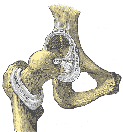
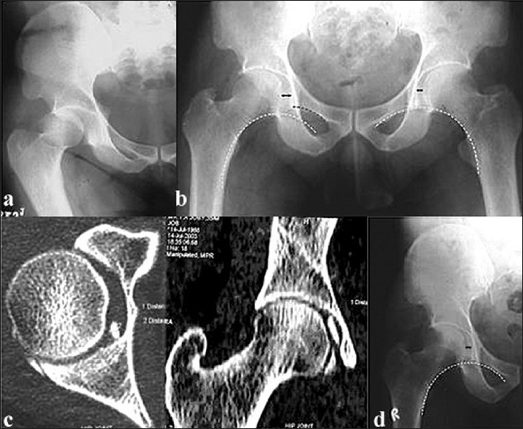
图 1-1 · 髋关节解剖(公有领域)Fig 1-1 · Hip anatomy (public domain) 观察点:右髋关节前方观(关节囊已大部切除):股骨头、髋臼、圆韧带清晰可见。正常时股骨头深坐于髋臼内,骨性匹配非常稳定——这正是脱位需要高能量暴力的解剖基础。Observation: anterior view of the right hip with the capsule largely removed: femoral head, acetabulum and ligamentum teres are visible. Normally the head sits deep in the socket with a highly stable bony match — the anatomic reason traumatic native dislocation usually follows high-energy trauma. 能证明:正常髋关节的骨性结构关系。不能证明:脱位方向、复位方法;1918 年解剖图谱,非影像。Shows: normal bony relationships of the hip. Does not show: dislocation direction or reduction technique; a 1918 anatomy plate, not imaging. 来源:Henry Gray《Anatomy of the Human Body》(1918) Plate 342,绘图 Henry Vandyke Carter,公有领域 · Wikimedia Commons 原页 · 独立图例,与本单元其他图非同一病例Source: Henry Gray, Anatomy of the Human Body (1918), Plate 342, illustrated by Henry Vandyke Carter, public domain · Wikimedia Commons file page · standalone figure, not the same case as other figures in this unit图 1-2 · 原生髋急性后脱位:复位不匹配与关节内游离体(CC BY)Fig 1-2 · Acute native posterior dislocation: incongruent reduction and loose body (CC BY) 观察点:44 岁男性车祸后右髋后脱位。(a) 脱位正位 X 线:股骨头向后上脱出髋臼;(b) 复位后正位:Shenton 线中断、内侧关节间隙增宽——复位不匹配,提示关节内嵌顿骨软骨游离体;(c) CT 证实关节内侧骨软骨碎片(后经开放取出);(d) 取出后复位恢复同心圆。本例说明复位后仍需检查关节匹配与残留骨片;不能用单例推导普遍检查策略。Observation: posterior dislocation of the right hip in a 44-year-old man after a crash. (a) Dislocated AP X-ray: femoral head displaced posterosuperiorly out of the acetabulum; (b) post-reduction AP: broken Shenton's line with widened medial joint space — incongruent reduction, suggesting an entrapped osteochondral loose body; (c) CT confirms a medial osteochondral fragment (later removed openly); (d) concentric reduction restored after removal. This case illustrates why congruence and residual fragments matter after reduction; a single case does not establish a universal imaging policy. 能证明:后脱位的 X 线表现;复位不匹配的征象(Shenton 线中断);CT 发现游离体的价值。不能证明:股骨头血供状态;坐骨神经状态(需查体)。Shows: the X-ray appearance of posterior dislocation; signs of incongruent reduction (broken Shenton's line); the value of CT for loose bodies. Does not show: femoral head blood supply; sciatic nerve status (needs examination). 来源:Karthik K 等,"Incongruent reduction following post-traumatic hip dislocations as an indicator of intra-articular loose bodies",《Indian Journal of Orthopaedics》2011,Fig. 1,CC BY 2.0(attribution license,许可说明)· PMC 原文 · 独立病例Source: Karthik K et al., "Incongruent reduction following post-traumatic hip dislocations as an indicator of intra-articular loose bodies", Indian Journal of Orthopaedics 2011, Fig. 1, CC BY 2.0 attribution license (license) · PMC article · standalone case拓展(折叠):假体脱位与少见脱位方向Extension (collapsed): prosthetic dislocation and uncommon directions
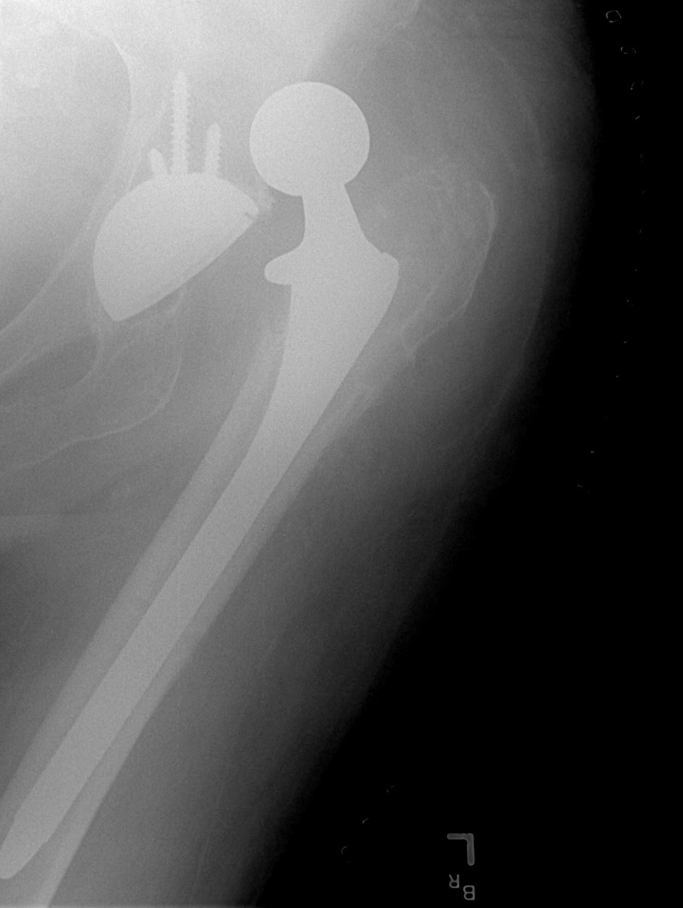
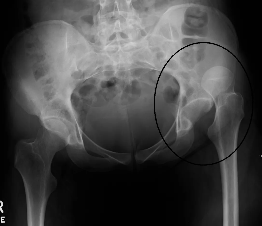
以下两图为鉴别拓展:假体脱位机制不同(体位/假体位置/软组织张力,可低能量发生),上方脱位少见。主课以原生髋急性后脱位(图 1-2)为准。The two figures below are extension material: prosthetic dislocation has a different mechanism (position/component placement/soft-tissue tension; can follow low energy), and superior dislocation is uncommon. The core teaching uses acute native posterior dislocation (Fig 1-2).
拓展图 1-A · 全髋假体脱位 X 线(CC BY 2.0)Extension Fig 1-A · Dislocated hip prosthesis X-ray (CC BY 2.0) 观察点:股骨柄假体头脱出髋臼杯、向上移位。假体脱位可见金属部件,机制常与体位、假体位置、软组织张力相关,可发生于低能量,复发风险更高——这正是课程把原生髋与假体脱位分开讨论的原因。Observation: the femoral stem head lies outside and above the acetabular cup. Metal components are visible; the mechanism often relates to position, component placement and soft-tissue tension, can follow low energy, and carries higher recurrence risk — why this course discusses native and prosthetic dislocations separately. 能证明:假体脱位的影像表现(假体头与臼杯分离)。不能证明:脱位原因(需结合体位史与假体位置评估)。Shows: the imaging appearance of prosthetic dislocation (head-cup separation). Does not show: the cause of dislocation (needs position history and component assessment). 来源:Bill Rhodes(Asheville),Wikimedia Commons,CC BY 2.0(许可)· 原页 · 独立病例Source: Bill Rhodes (Asheville), via Wikimedia Commons, CC BY 2.0 (license) · file page · standalone case拓展图 1-B · 上方脱位 X 线(CC BY-SA 3.0,少见方向)Extension Fig 1-B · Superior dislocation X-ray (CC BY-SA 3.0, uncommon direction) 观察点:骨盆正位:左侧(图片右侧)股骨头向上移位、髋臼空虚,为创伤后上方脱位(上传者原标注圈出)。上方脱位少见,临床最常见的是后脱位(约90%,见图 1-2);读片要点相同——看股骨头是否位于髋臼内。Observation: AP pelvis: the left femoral head (image right) is displaced superiorly with an empty acetabulum — post-traumatic superior dislocation (circled by the uploader). Superior dislocation is uncommon; posterior dislocation (~90%, see Fig 1-2) is most common clinically; the reading point is the same — is the head inside the acetabulum? 能证明:脱位时"髋臼空虚、股骨头异位"的 X 线征象。不能证明:脱位方向的分布(单例不能代表 90% 后脱位);不能显示关节囊撕裂与坐骨神经状态。Shows: the "empty acetabulum, displaced head" sign of dislocation. Does not show: the distribution of dislocation directions (one case cannot represent ~90% posterior); cannot show capsular tearing or sciatic nerve status. 来源:James Heilman, MD,Wikimedia Commons,CC BY-SA 3.0(许可)· 原页 · 独立病例Source: James Heilman, MD, via Wikimedia Commons, CC BY-SA 3.0 (license) · file page · standalone case
AO 原则如何适用How AO principles apply
解剖复位:脱位本身要求尽快闭合复位,恢复关节的同心圆关系。Anatomic reduction: the dislocation itself demands urgent closed reduction to restore a concentric joint.
稳定固定:合并的髋臼、股骨头骨折,按骨折的 AO 原则固定。Stable fixation: associated acetabular/femoral-head fractures are fixed per AO fracture principles.
早期活动:在关节稳定、骨折允许的前提下尽早活动。Early mobilization: as soon as joint stability and fractures allow.
复位操作的基本要求(原则层面)Basic requirements for reduction (principles)
在充分镇静/麻醉和肌松下进行,避免暴力。Perform under adequate sedation/anesthesia with muscle relaxation; avoid force.
复位后立即复查神经血管状态和影像。Immediately recheck neurovascular status and imaging after reduction.
本课程不演示具体复位手法。Specific reduction maneuvers are not demonstrated in this course.
治疗选择与限制Treatment options & limitations
尽快复位,不存在可等待的"安全时间窗"。未复位的脱位持续威胁股骨头血供,延迟增加缺血坏死风险——"越快越好"不是"6小时内安全"。Reduce ASAP — there is no safe waiting window. An unreduced dislocation keeps threatening head blood supply; delay raises AVN risk. "As soon as possible" does not mean "safe within 6 hours".
原生髋:无阻碍闭合复位的合并损伤时,通常先尝试闭合复位;复位失败、复位后不稳定或合并需固定的骨折 → 切开复位。Native hip: usually attempt closed reduction when associated injuries do not preclude it; failed reduction, post-reduction instability, or fractures needing fixation → open reduction.
假体脱位:多数可闭合复位;复位后必须评估脱位原因(体位习惯、假体位置、软组织张力),并做防脱位宣教。Prosthetic dislocation: usually reducible closed; afterwards the cause (habitual positions, component placement, soft-tissue tension) must be assessed, with dislocation-precaution education.
限制:合并股骨颈骨折时,闭合复位可能加重骨折移位,需按脱位合并骨折的联合损伤原则处理(见来源)。Limitation: with a femoral neck fracture, closed reduction may worsen displacement — manage as a combined dislocation-fracture injury (see sources).
并发症 / 随访Complications / follow-up
股骨头缺血坏死(AVN):随访中用影像监测,坏死可在伤后一段时间内才显现。Avascular necrosis (AVN) of the femoral head: monitor with imaging; it may declare itself after an interval.
坐骨神经损伤:多数可逐渐恢复,需定期随访神经功能。Sciatic nerve injury: often recovers gradually; follow neurologic function.
假体再脱位:按手术入路、假体与稳定性进行个体化体位防护宣教。Prosthetic re-dislocation: teach position precautions per surgeon advice.
病例自测Case quiz
Q1 · 28岁男性,车祸后右下肢呈屈曲、内收、内旋、缩短畸形,足背感觉减退、足下垂。最可能的诊断和最需要警惕的神经是?(合成教学病例)Q1 · A 28-year-old man after a car crash: right leg flexed, adducted, internally rotated and shortened, with dorsal foot sensory loss and foot drop. Most likely diagnosis and nerve at risk? (synthetic teaching case)
回答正确 ✓Correct ✓
再想想 ✗Try again ✗
答案:B。Answer: B.屈曲内收内旋缩短是后脱位典型体位;后脱位最易伤及坐骨神经,其中腓总神经支最脆弱,表现为足下垂。Flexed, adducted, internally rotated and shortened is classic for posterior dislocation; the sciatic nerve — especially its peroneal division — is most vulnerable, causing foot drop.
Q2 · 关于髋关节脱位复位时机的说法,正确的是?Q2 · Which statement about timing of hip reduction is correct?
回答正确 ✓Correct ✓
再想想 ✗Try again ✗
答案:B。Answer: B.未复位的脱位持续威胁股骨头血供。"6小时"不是可以放心等待的窗口,而是越快越好。CT是复位后的常规检查,不是复位的前提。An unreduced dislocation keeps threatening head blood supply. "6 hours" is not a window to wait inside — sooner is better. CT is routine after reduction, not a prerequisite.
来源与媒体许可Sources & media licensing
AO Surgery Reference — Split fractures of the femoral head (definition) 定位:未复位脱位是急症(威胁股骨头血供、可压迫重要神经);后脱位约90%、前脱位约10%。访问日期 2026-10-04。Relevance: unreduced dislocation is an emergency (threatens head blood supply, may compress major nerves); ~90% posterior, ~10% anterior. Accessed 2026-10-04.
媒体来源:图 1-1 为公有领域解剖图(Gray's Anatomy 1918);图 1-2 CC BY(Karthik K 等,《Indian Journal of Orthopaedics》2011,PMC3004076);拓展图 1-A CC BY 2.0(Bill Rhodes)、拓展图 1-B CC BY-SA 3.0(James Heilman, MD),均随包附带、原页与许可见各图注;AO Surgery Reference 为外部来源链接,仅引用未复制其图文与视频,内容版权归 AO Foundation 所有(AO 未提供公开再发布许可)。Media sources: Fig 1-1 is a public-domain anatomy plate (Gray's Anatomy, 1918); Fig 1-2 CC BY (Karthik K et al., Indian Journal of Orthopaedics 2011, PMC3004076); Extension Figs 1-A CC BY 2.0 (Bill Rhodes) and 1-B CC BY-SA 3.0 (James Heilman, MD), all bundled with source pages and licenses in their captions; AO Surgery Reference is an external source linked for reference only — no content copied; copyright remains with the AO Foundation (AO offers no open redistribution license).
远端 33常累及膝关节面;双峰分布——年轻人高能量、老年人低能量骨质疏松。Distal 33Often involves the knee joint; bimodal — high energy in the young, low-energy osteoporotic in the elderly.
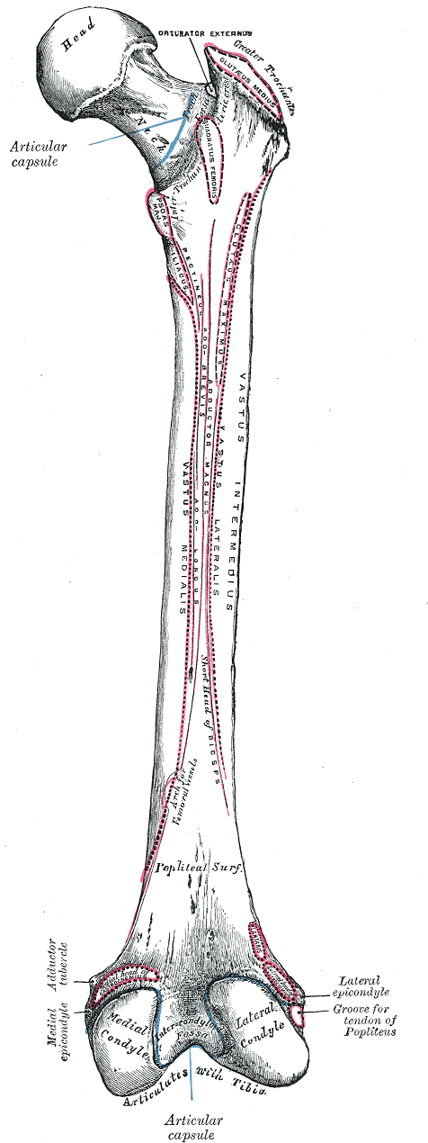
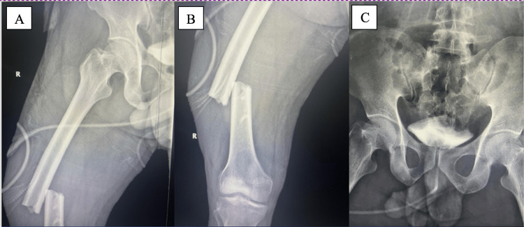
图 2-1 · 股骨解剖与三段划分(公有领域)Fig 2-1 · Femur anatomy and three segments (public domain) 观察点:右股骨后面观:股骨头、股骨颈、大小转子、骨干、内外侧髁。对照 AO/OTA:近端 31(头颈转子区)、骨干 32、远端 33(髁区)——三段的血供风险、固定方式与康复逻辑都不同。Observation: posterior view of the right femur: head, neck, greater/lesser trochanters, shaft, condyles. Maps to AO/OTA: proximal 31, shaft 32, distal 33 — blood-supply risk, fixation and rehab logic differ by segment. 能证明:三段的解剖分界。不能证明:骨折本身;1918 年解剖图谱,非影像。Shows: the anatomic boundaries of the three segments. Does not show: any fracture; a 1918 anatomy plate, not imaging. 来源:Henry Gray《Anatomy of the Human Body》(1918) Plate 245,绘图 Henry Vandyke Carter,公有领域 · Wikimedia Commons 原页 · 独立图例Source: Henry Gray, Anatomy of the Human Body (1918), Plate 245, illustrated by Henry Vandyke Carter, public domain · Wikimedia Commons file page · standalone figure图 2-2 · 创伤性股骨干骨折(CC BY)Fig 2-2 · Traumatic femoral shaft fracture (CC BY) 观察点:35 岁男性、100 km/h 正面撞击车祸后右股骨中段闭合骨折。(A)(B) 股骨正位 X 线:骨干中段骨折、断端明显移位;(C) 骨盆正位(术前评估的一部分)。这是典型的高能量创伤性股骨干骨折——对应课程"骨干 32 多为高能量暴力,肌肉牵拉使骨折端明显移位"。Observation: closed right midshaft femoral fracture in a 35-year-old man after a frontal crash at ~100 km/h. (A)(B) AP femur X-rays: midshaft fracture with marked displacement; (C) AP pelvis (part of preoperative assessment). This is a typical high-energy traumatic femoral shaft fracture — matching the course point "32 shaft fractures are mostly high-energy, with muscle pull displacing the fragments". 能证明:高能量股骨干骨折的 X 线表现(断端明显移位)。不能证明:同侧股骨颈隐匿骨折(需加拍髋部影像);固定方式(本图为术前片)。Shows: the X-ray appearance of a high-energy shaft fracture (marked displacement). Does not show: an occult ipsilateral femoral neck fracture (needs dedicated hip imaging); the fixation method (preoperative film). 来源:Alzahrani JA 等,Cureus 2023,Fig. 1,CC BY(原文未标版本;许可)· PMC 原文 · 独立病例Source: Alzahrani JA et al., Cureus 2023, Fig. 1, CC BY (version unstated in source; license) · PMC article · standalone case拓展(折叠):非典型股骨骨折(双膦酸盐相关)Extension (collapsed): atypical femoral fracture (bisphosphonate-associated)
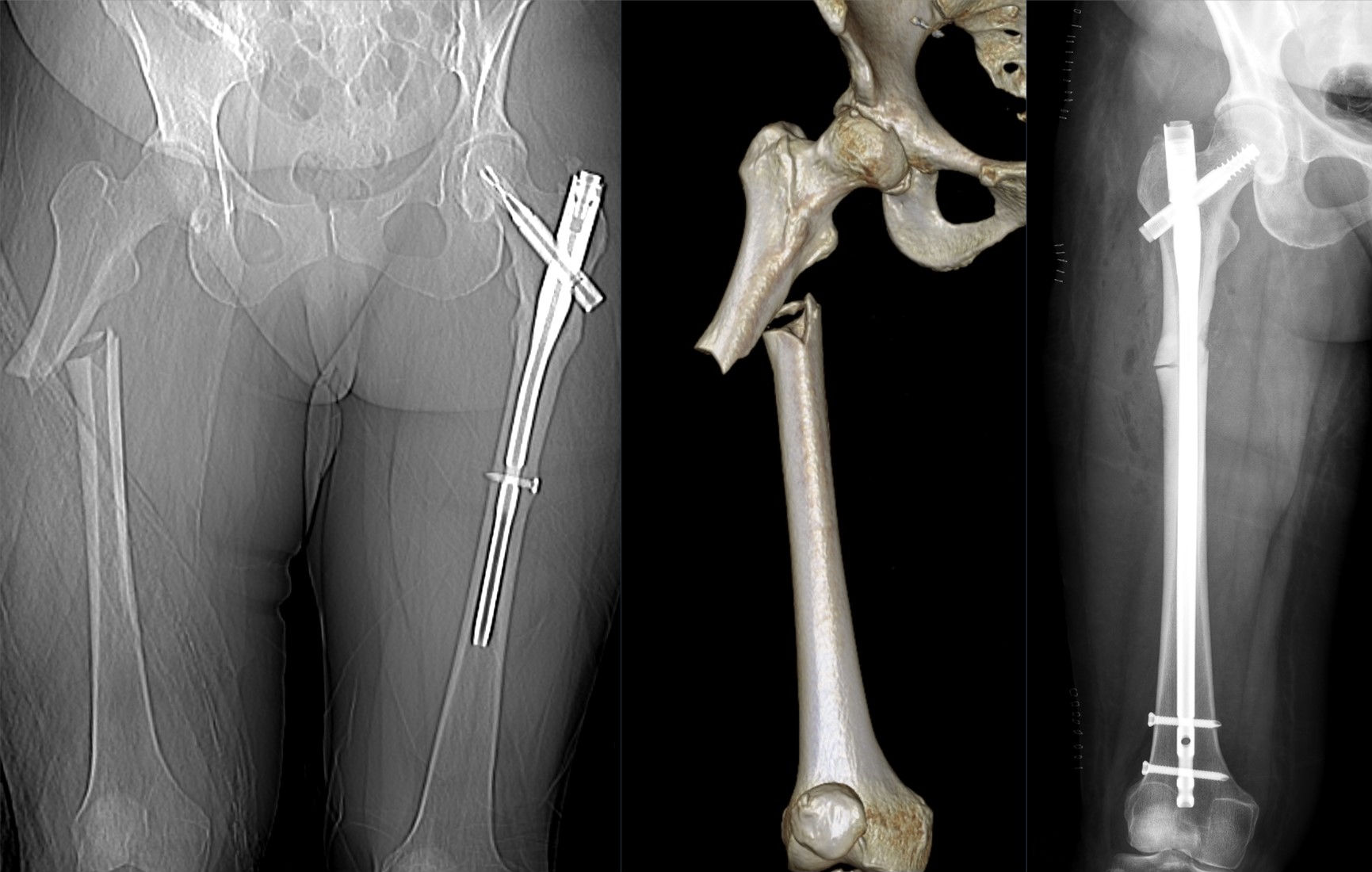
下图为鉴别拓展:长期双膦酸盐治疗相关非典型股骨干骨折可发生于低能量、骨折线形态不同(横形、骨皮质增厚)。不能以此代表高能量创伤——主课以创伤性股骨干骨折(图 2-2)为准。The figure below is extension material: bisphosphonate-associated atypical femoral shaft fractures can follow low energy and look different (transverse line, cortical thickening). It cannot represent high-energy trauma — the core teaching uses traumatic femoral shaft fracture (Fig 2-2).
拓展图 2-A · 非典型股骨干骨折(CC BY-SA 4.0,双膦酸盐相关)Extension Fig 2-A · Atypical femoral shaft fracture (CC BY-SA 4.0, bisphosphonate-associated) 观察点:组合图(CT 定位像 + 三维重建 + 术后 X 线):右侧股骨近端骨干横形骨折,骨折线基本横贯,外侧皮质原有增厚(既往应力性改变);术后 X 线示髓内钉贯穿骨折端。本例为双膦酸盐相关的非典型骨折(低能量),用于观察横形形态与髓内钉固定;创伤性股骨干骨折多为高能量暴力(见图 2-2)。Observation: composite (CT scout + 3D reconstruction + post-op X-ray): transverse fracture of the proximal femoral shaft with pre-existing lateral cortical thickening (prior stress change); the post-op film shows an intramedullary nail spanning the fracture. This is a bisphosphonate-related atypical fracture (low energy), shown here for the transverse morphology and nailing; traumatic shaft fractures are usually high energy (see Fig 2-2). 能证明:横形骨折形态、髓内钉的固定方式。不能证明:高能量创伤的典型表现;不能代表所有股骨干骨折的病因。Shows: transverse fracture morphology and intramedullary nailing. Does not show: typical high-energy trauma; does not represent all causes of femoral shaft fracture. 来源:Hellerhoff,Wikimedia Commons,CC BY-SA 4.0(许可)· 原页 · 独立病例Source: Hellerhoff, via Wikimedia Commons, CC BY-SA 4.0 (license) · file page · standalone case
初评和红旗Initial assessment & red flags
股骨干骨折出血量大,按多发伤流程评估循环;老年人近端骨折重点评估全身合并症。Shaft fractures bleed significantly — assess circulation as in polytrauma; in elderly proximal fractures, assess comorbidities.
红旗Red flags
同侧股骨颈隐匿骨折:股骨干骨折必须加拍髋部,以免漏诊。Occult ipsilateral femoral-neck fracture: shaft fractures need hip imaging to avoid missing it.
开放骨折:尽早清创覆盖,抗生素与 tetanus 按规范。Open fracture: early debridement and coverage; antibiotics and tetanus per protocol.
远端骨折:查足背/胫后动脉搏动与末梢灌注(皮色、皮温、毛细血管再充盈),警惕血管损伤;注意脉搏存在不能完全排除血管伤;查腓总神经。Distal fractures: check dorsalis pedis/posterior tibial pulses and distal perfusion (color, warmth, capillary refill); note a present pulse does not fully exclude vascular injury; check the peroneal nerve.
多发伤:股骨干骨折常合并胸、骨盆损伤。Polytrauma: shaft fractures often accompany chest/pelvic injuries.
影像 / 分类(三段)Imaging / classification (by segment)
近端:骨盆正位 + 患髋正侧位;Garden 描述股骨颈移位程度(描述性)。Proximal: AP pelvis + hip views; Garden describes neck displacement (descriptive).
骨干:全长股骨正侧位(含髋、膝关节);AO/OTA 32 简单 / 楔形 / 复杂(描述性)。Shaft: full-length femur AP/lateral including hip and knee; AO/OTA 32 simple / wedge / complex (descriptive).
分类 ≠ 手术指征Classification ≠ indication:分类描述骨折形态;是否手术、用什么固定,还取决于年龄、骨质量、软组织与全身情况。: classification describes morphology; whether and how to fix also depends on age, bone quality, soft tissue and overall condition.
AO 原则如何适用(三段)How AO principles apply (by segment)
近端:解剖复位 + 稳定固定,核心是保护股骨头血供;移位股骨颈骨折在老年患者中,关节置换是讨论选项之一。Proximal: anatomic reduction + stable fixation, centered on protecting head blood supply; arthroplasty is one discussed option for displaced neck fractures in the elderly.
骨干:髓内钉是成人常规选择——闭合穿钉、微创、保护骨折端血肿,允许早期活动。Shaft: intramedullary nailing is routine in adults — closed, minimally invasive, preserves fracture hematoma, allows early mobilization.
远端:关节面解剖复位 + 稳定固定 + 早期活动;干骺端粉碎与骨质疏松是难点。Distal: anatomic articular reduction + stable fixation + early motion; metaphyseal comminution and osteoporosis are the challenges.
骨干:顺行 vs 逆行髓内钉(原则层面)Shaft: antegrade vs retrograde nailing (principles)
顺行钉是常用入路;逆行钉对远端 1/3 骨折有优势,两者总体疗效相近(见来源)。Antegrade nailing is commonly used; retrograde nails suit distal-third fractures, with similar overall outcomes (see sources).
微创钢板(MIPO)可减少肌肉剥离与血运破坏,按骨折部位选择入路。Minimally invasive plating reduces muscle stripping and vascular compromise; approach varies by fracture location.
近端:选项包括空心钉、滑动髋螺钉(DHS)、髓内钉(转子间)、关节置换。限制:囊内骨折的缺血坏死风险、骨质疏松、患者生理储备——方案必须个体化。Proximal: options include cannulated screws, sliding hip screw (DHS), cephalomedullary nailing (intertrochanteric), arthroplasty. Limits: AVN risk in intracapsular fractures, osteoporosis, physiologic reserve — decisions must be individualized.
骨干:髓内钉为常规。限制:开放骨折、多发伤(可先损伤控制外固定)、严重粉碎。非手术用于无手术条件、不能耐受手术等个体情况(AO Surgery Reference 明确)。Shaft: nailing is routine. Limits: open fractures, polytrauma (damage-control ex-fix first), severe comminution. Nonoperative for lack of surgical capacity, inability to tolerate surgery, or other individual factors (stated in AO Surgery Reference).
远端:锁定钢板、逆行髓内钉。限制:严重关节粉碎、开放伤、血管损伤需先处理血管。Distal: locking plates, retrograde nailing. Limits: severe articular comminution, open injury, vascular injury requiring vascular care first.
随访:按计划复查影像、渐进负重、功能康复;内固定激惹可在骨折愈合后、经专科评估考虑取出(无统一时间标准)。Follow up with scheduled imaging, progressive weight bearing and rehab; symptomatic implants may be removed after healing, following specialist assessment (no universal timing).
病例自测Case quiz
Q1 · 24岁男性,车祸致右股骨干横形骨折,闭合。按 AO Surgery Reference,常规治疗选择是?(合成教学病例)Q1 · A 24-year-old man with a closed transverse femoral shaft fracture after a crash. Per AO Surgery Reference, the routine choice is? (synthetic teaching case)
回答正确 ✓Correct ✓
再想想 ✗Try again ✗
答案:C。Answer: C.成人股骨干骨折通常手术,髓内钉为常规;非手术用于无手术条件、不能耐受手术等个体情况。Adult shaft fractures are normally treated operatively with nailing; nonoperative care is for lack of surgical capacity, inability to tolerate surgery, or other individual factors.
Q2 · 股骨干骨折的影像检查中,最容易漏诊的合并伤是?Q2 · In femoral shaft fractures, the most easily missed associated injury on imaging is?
回答正确 ✓Correct ✓
再想想 ✗Try again ✗
答案:A。Answer: A.这就是要求拍全长股骨(含髋、膝)的原因:同侧股骨颈隐匿骨折一旦漏诊后果严重。This is why full-length femur films including hip and knee are required: a missed neck fracture has serious consequences.
Q3 · 远端股骨骨折,CT最重要的价值是?Q3 · In distal femur fractures, the most important value of CT is?
回答正确 ✓Correct ✓
再想想 ✗Try again ✗
答案:B。Answer: B.相当一部分远端股骨骨折有平片看不见的关节内延伸,CT 决定你是否需要处理关节面。A substantial share of distal femur fractures have occult intra-articular extension; CT determines whether the joint surface needs addressing.
媒体来源:图 2-1 为公有领域解剖图(Gray's Anatomy 1918);图 2-2 CC BY(Alzahrani JA 等,Cureus 2023,PMC10440397);拓展图 2-A CC BY-SA 4.0(Hellerhoff),均随包附带、原页与许可见各图注;AO Surgery Reference 为外部来源链接,仅引用未复制其图文与视频,内容版权归 AO Foundation 所有(AO 未提供公开再发布许可)。Media sources: Fig 2-1 is a public-domain anatomy plate (Gray's Anatomy, 1918); Fig 2-2 CC BY (Alzahrani JA et al., Cureus 2023, PMC10440397); Extension Fig 2-A CC BY-SA 4.0 (Hellerhoff), all bundled with source pages and licenses in their captions; AO Surgery Reference is an external source linked for reference only — no content copied; copyright remains with the AO Foundation (AO offers no open redistribution license).
3髌骨骨折Patella fracture
伸膝装置是核心——先判断装置断没断,再谈固定方式The extensor mechanism is central — assess it first, then discuss fixation
官方影像资源(AO Surgery Reference,站内查看)Official imaging (AO Surgery Reference, view on-site)
直接暴力(跌倒膝着地、仪表盘撞击):多为粉碎骨折。Direct trauma (fall onto the knee, dashboard impact): often comminuted.
间接暴力(屈膝位股四头肌猛烈收缩):多为横形骨折,常伴分离。Indirect force (violent quadriceps contraction with knee flexed): often transverse, frequently displaced.
髌骨骨折常合并髌股关节面损伤。Patellar fractures often injure the patellofemoral articular surface.
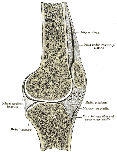
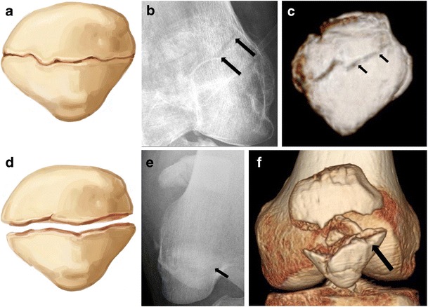
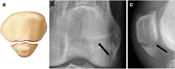
图 3-1 · 膝关节矢状切面与伸膝装置(公有领域)Fig 3-1 · Knee sagittal section and extensor mechanism (public domain) 观察点:右膝关节矢状切面:股骨、髌骨(patella)、髌韧带(ligamentum patellae)、胫骨、半月板。伸膝装置是一条链:股四头肌腱(上)→ 髌骨 → 髌韧带 → 胫骨结节——任何一环断裂,伸膝都受影响,这正是"先判断装置断没断,再谈固定方式"的解剖依据。Observation: sagittal section of the right knee: femur, patella, patellar ligament, tibia, meniscus. The extensor mechanism is a chain — quadriceps tendon → patella → patellar ligament → tibial tuberosity; a break anywhere impairs knee extension — the anatomic basis for "assess the mechanism first, then discuss fixation". 能证明:伸膝装置的解剖连续关系。不能证明:骨折;1918 年解剖图谱,非影像。Shows: the anatomic continuity of the extensor mechanism. Does not show: any fracture; a 1918 anatomy plate, not imaging. 来源:Henry Gray《Anatomy of the Human Body》(1918) Plate 350,绘图 Henry Vandyke Carter,公有领域 · Wikimedia Commons 原页 · 独立图例Source: Henry Gray, Anatomy of the Human Body (1918), Plate 350, illustrated by Henry Vandyke Carter, public domain · Wikimedia Commons file page · standalone figure图 3-2 · 横行髌骨骨折:无移位 vs 移位(CC BY 4.0)Fig 3-2 · Transverse patellar fractures: non-displaced vs displaced (CC BY 4.0) 观察点:两名年轻成人的横行骨折对照。上排(a–c):无移位横行骨折——示意图、正位 X 线、CT 三维重建,骨折线横贯髌骨体部;下排(d–f):移位横行骨折——两骨折端分离。注意:上排与下排是两名不同患者,不是同一病例的时序。移位者支持带损伤风险更高——对应课程"分离的横形骨折多伴装置断裂"。Observation: transverse fractures in two young adults compared. Top row (a–c): non-displaced transverse fracture — drawing, AP radiograph, 3D CT reconstruction, fracture line across the patellar body; bottom row (d–f): displaced transverse fracture with fragment separation. Note: the two rows are two different patients, not one case over time. Displaced fractures carry higher retinacular injury risk — matching the course point that displaced transverse fractures usually mean a disrupted mechanism. 能证明:横行骨折的形态(无移位 vs 移位)、X 线与 CT 的互补。不能证明:伸膝装置查体所见;两名患者均为年轻成人,不能代表老年骨质疏松骨折。Shows: transverse fracture morphology (non-displaced vs displaced) and the complementary roles of X-ray and CT. Does not show: physical-exam findings of the extensor mechanism; both patients are young adults, not representative of elderly osteoporotic fractures. 来源:Jarraya M 等,"Imaging of patellar fractures",《Insights into Imaging》2017,Fig. 3,CC BY 4.0(许可)· PMC 原文 · 两名独立患者Source: Jarraya M et al., "Imaging of patellar fractures", Insights into Imaging 2017, Fig. 3, CC BY 4.0 (license) · PMC article · two standalone patients图 3-3 · 髌骨下极骨折(CC BY 4.0)Fig 3-3 · Inferior pole patellar fracture (CC BY 4.0) 观察点:39 岁男性髌骨下极横行骨折:示意图、正位与侧位 X 线。本图展示下极骨折线;是否合并髌腱损伤、髌骨高度是否异常,应结合完整侧位片、查体及必要的进一步检查,不能只凭此图确定。Observation: transverse fracture of the lower pole in a 39-year-old man: drawing, AP and lateral radiographs. The image shows the lower-pole fracture line. Tendon integrity and patellar height require the full lateral view, examination and further investigation when indicated; this image alone is insufficient. 能证明:下极骨折的 X 线表现。不能证明:髌腱是否完全断裂(需超声/MRI);与图 3-2 非同一患者。Shows: the X-ray appearance of an inferior pole fracture. Does not show: whether the patellar tendon is fully ruptured (needs ultrasound/MRI); not the same patient as Fig 3-2. 来源:Jarraya M 等,"Imaging of patellar fractures",《Insights into Imaging》2017,Fig. 4,CC BY 4.0(许可)· PMC 原文 · 独立病例Source: Jarraya M et al., "Imaging of patellar fractures", Insights into Imaging 2017, Fig. 4, CC BY 4.0 (license) · PMC article · standalone case
初评和红旗Initial assessment & red flags
关键检查是伸膝装置:让患者做主动直腿抬高;能完成 → 装置可能完整;出现伸膝滞后 → 提示伸膝装置损伤(疼痛抑制也可能影响,需综合评估)。The key exam is the extensor mechanism: ask for an active straight-leg raise; able to do it → mechanism may be intact; extensor lag → suggests extensor injury (pain inhibition can also affect it; assess comprehensively).
触诊有无骨缺损(台阶感),观察关节积血。Palpate for a bony defect (step); look for hemarthrosis.
注意:髌旁支持带完整时,患者仍可能伸直膝盖,不能单凭"能伸直"排除骨折。Note: with intact retinacula, patients may still extend the knee — ability to extend does not rule out fracture.
红旗Red flags
开放性骨折:膝前位置表浅,开放并不少见 → 按开放骨折处理。Open fracture: the anterior knee is subcutaneous, so open injuries are not rare → manage as open fracture.
不要把髌前滑囊损伤误认为骨折,也不要漏掉开放伤的分级。Don't confuse prepatellar bursal injury with fracture; don't under-grade open injuries.
检查不确定时怎么办?What if the exam is uncertain?
对伸膝装置是否断裂有疑问时,可做超声或 MRI 明确股四头肌腱与髌腱情况(见来源)。When disruption is uncertain, ultrasound or MRI can clarify quadriceps/patellar tendon status (see sources).
影像 / 分类Imaging / classification
膝正侧位 + 髌骨轴位(skyline);侧位最能显示分离程度。Knee AP/lateral + skyline views; the lateral best shows displacement.
CT:评估粉碎程度与关节面台阶。CT: assess comminution and articular step-off.
AO/OTA 34:关节外 / 部分关节内 / 完全关节内(描述性);横形骨折伴分离通常意味着伸膝装置断裂。AO/OTA 34: extra-articular / partial-articular / complete-articular (descriptive); transverse fractures with separation usually mean a disrupted mechanism.
分类 ≠ 手术指征Classification ≠ indication:分类描述形态;真正分流治疗的是伸膝装置是否完整和移位程度。: classification describes morphology; what truly triages treatment is whether the extensor mechanism is intact plus displacement.
AO 原则如何适用How AO principles apply
恢复伸膝装置的连续性是第一目标,其次是关节面平整。Restoring extensor continuity is the first goal, then a smooth articular surface.
稳定固定允许早期活动,对抗膝僵硬。Stable fixation permits early motion, fighting knee stiffness.
髌骨血供相对脆弱、软组织覆盖薄,骨折与手术都可能进一步影响血运,手术要爱护软组织。The patella has relatively tenuous blood supply and thin soft-tissue cover, and both fracture and surgery can compromise it further — handle soft tissues gently.
治疗选择与限制Treatment options & limitations
伸膝装置完整 + 无移位/轻微移位:可考虑非手术——伸直位支具固定 4–6 周,允许负重(见来源)。Intact mechanism + undisplaced/minimally displaced: nonoperative may be considered — extension bracing 4–6 weeks with weight bearing allowed (see sources).
装置断裂或明显移位:手术重建——张力带、螺钉等,恢复装置连续与关节面。Disrupted mechanism or marked displacement: surgical reconstruction — tension band, screws, etc., restoring continuity and the joint surface.
限制:严重粉碎无法重建、患者全身情况不允许手术、开放伤软组织条件差——方案个体化。Limits: unreconstructable comminution, unfit patients, poor soft tissue in open injuries — individualize.
并发症 / 随访Complications / follow-up
膝僵硬:最常见,早期活动是预防关键。Knee stiffness: most common; early motion is key prevention.
创伤性髌股关节炎:关节面损伤越重风险越高。Patellofemoral arthritis: worse with greater articular injury.
内固定激惹/失败:髌前软组织薄,内植物激惹常见。Implant irritation/failure: common given thin prepatellar cover.
Q1 · 45岁女性,跌倒后右膝肿痛,X线示髌骨横形骨折、分离约8mm。查体最能决定治疗方向的检查是?(合成教学病例)Q1 · A 45-year-old woman fell onto her knee; X-ray shows a transverse patella fracture with ~8 mm separation. The exam finding that most directs treatment is? (synthetic teaching case)
回答正确 ✓Correct ✓
再想想 ✗Try again ✗
答案:B。Answer: B.伸膝装置是否完整是分流治疗的关键:分离的横形骨折多伴装置断裂,需手术重建。Extensor integrity triages treatment: displaced transverse fractures usually mean a disrupted mechanism needing reconstruction.
Q2 · 关于髌骨骨折的说法,正确的是?Q2 · Which statement about patella fractures is correct?
回答正确 ✓Correct ✓
再想想 ✗Try again ✗
答案:A。Answer: A.辅助伸膝结构(内外侧髌旁支持带)完整时可代偿部分伸膝功能;膝前位置表浅,开放伤并不少见。Intact medial/lateral retinacula can compensate partly; the subcutaneous anterior knee makes open injuries not rare.
来源与媒体许可Sources & media licensing
AO Surgery Reference — Patient examination (patella) 定位:伸膝装置检查(直腿抬高、支持带代偿、开放伤评估)。访问日期 2026-10-04。Relevance: extensor mechanism exam (straight-leg raise, retinacular compensation, open-injury assessment). Accessed 2026-10-04.
媒体来源:图 3-1 为公有领域解剖图(Gray's Anatomy 1918);图 3-2、图 3-3 为 CC BY 4.0(Jarraya M 等,《Insights into Imaging》2017,PMC5265199),随包附带、原页与许可见图注;AO Surgery Reference 为外部来源链接,仅引用未复制其图文与视频,内容版权归 AO Foundation 所有(AO 未提供公开再发布许可)。Media sources: Fig 3-1 is a public-domain anatomy plate (Gray's Anatomy, 1918); Figs 3-2 and 3-3 CC BY 4.0 (Jarraya M et al., Insights into Imaging 2017, PMC5265199), bundled with source pages and licenses in their captions; AO Surgery Reference is an external source linked for reference only — no content copied; copyright remains with the AO Foundation (AO offers no open redistribution license).
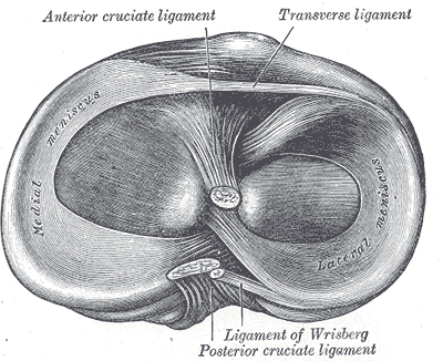
胫骨平台:内侧大而凹、外侧小而凸,后倾约10°;内侧承担更多负荷。The plateau: medial is large and concave, lateral small and convex, with ~10° posterior slope; the medial side bears more load.
机制:轴向暴力 + 内外翻应力。年轻人多为高能量(车祸、坠落);老年人骨质疏松,低能量跌倒即可骨折。Mechanism: axial load + varus/valgus stress. Young patients: high energy (traffic, falls); elderly osteoporotic patients: low-energy falls suffice.
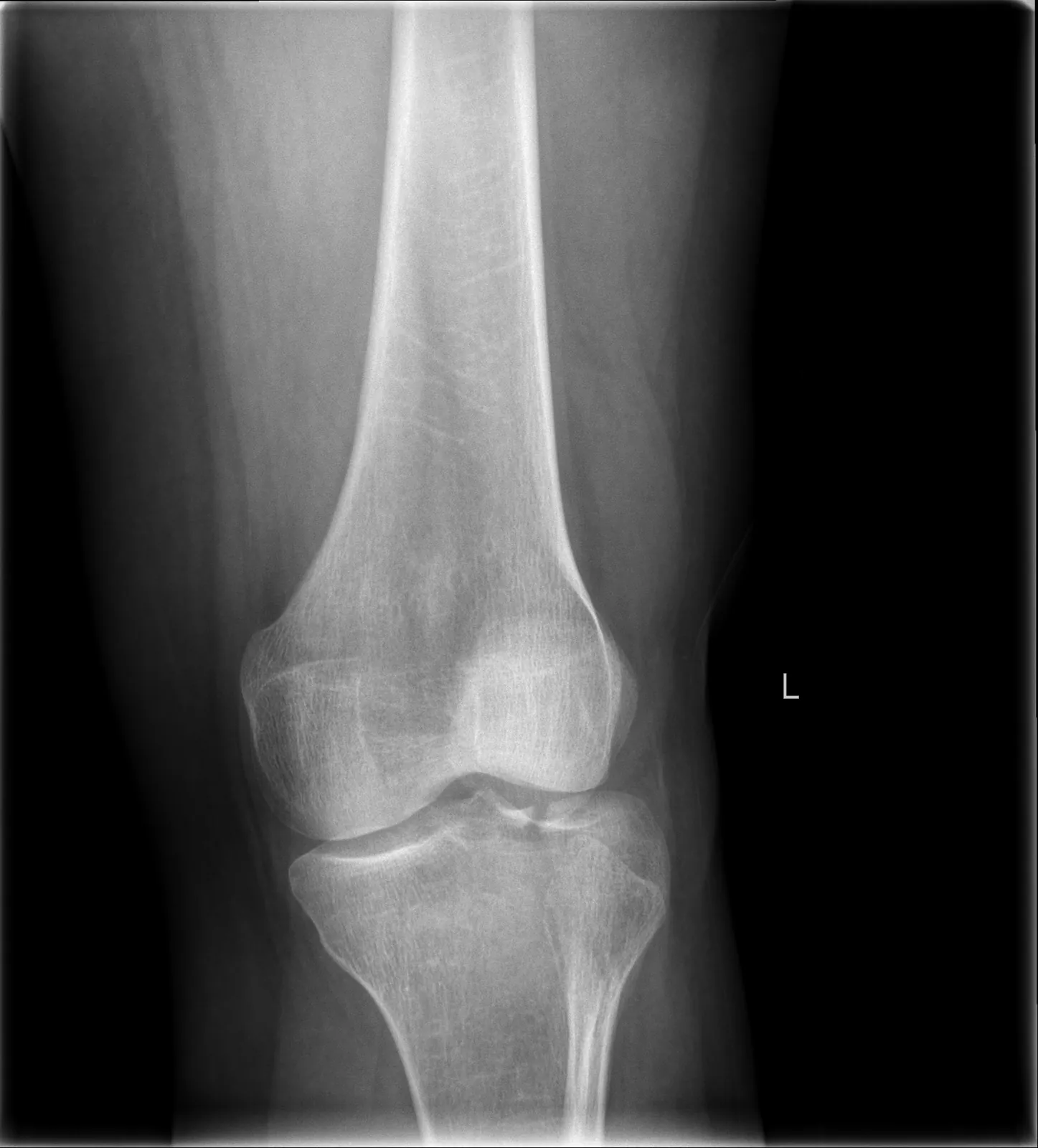
典型形态:外侧劈裂塌陷最常见;高能量可致双髁粉碎。Typical pattern: lateral split-depression is most common; high energy can shatter both condyles.
图 4-1 · 胫骨上端俯视:内外侧形态差异(公有领域)Fig 4-1 · Tibial head from above: medial–lateral difference (public domain) 观察点:右胫骨上端俯视:内侧半月板大而 C 形、外侧半月板小而 O 形;前后交叉韧带附着于髁间区。对应平台形态:内侧平台大而凹、外侧小而凸——外侧骨质相对薄弱,劈裂塌陷好发于外侧。理解这个形态差异,才能读懂图 4-2 的骨折。Observation: superior view of the right tibial head: medial meniscus large and C-shaped, lateral small and O-shaped; cruciate ligaments attach in the intercondylar area. Matches plateau morphology — medial large/concave, lateral small/convex — the lateral side is relatively weaker, where split-depression typically occurs. Grasping this difference is key to reading Fig 4-2. 能证明:内外侧平台的形态差异。不能证明:骨折;1918 年解剖图谱,非影像。Shows: the medial–lateral morphologic difference. Does not show: any fracture; a 1918 anatomy plate, not imaging. 来源:Henry Gray《Anatomy of the Human Body》(1918) Plate 349,绘图 Henry Vandyke Carter,公有领域 · Wikimedia Commons 原页 · 独立图例Source: Henry Gray, Anatomy of the Human Body (1918), Plate 349, illustrated by Henry Vandyke Carter, public domain · Wikimedia Commons file page · standalone figure图 4-2 · 外侧平台劈裂塌陷 X 线(CC BY-SA 4.0)Fig 4-2 · Lateral plateau split-depression X-ray (CC BY-SA 4.0) 观察点:膝关节正位 X 线(L 标记):外侧胫骨平台塌陷,关节面较内侧下沉、失去平整。对应"外侧劈裂塌陷"——平台骨折最常见的形态之一,与图 4-1 的外侧薄弱形态相呼应。Observation: AP knee X-ray (L marker): depressed lateral tibial plateau with loss of articular flatness compared with the medial side. Typical lateral split-depression — one of the most common plateau patterns, echoing the weaker lateral morphology in Fig 4-1. 能证明:外侧平台塌陷的 X 线表现。不能证明:塌陷的精确深度(需 CT);软组织状态;与图 4-1 非同一来源。Shows: the X-ray appearance of lateral plateau depression. Does not show: the exact depth of depression (needs CT); soft-tissue status; not the same source as Fig 4-1. 来源:LottieLattes,Wikimedia Commons,CC BY-SA 4.0(许可)· 原页 · 独立病例Source: LottieLattes, via Wikimedia Commons, CC BY-SA 4.0 (license) · file page · standalone case
初评和红旗Initial assessment & red flags
软组织是第一位的:记录肿胀程度、水疱(血疱/张力性水疱)、皮肤张力、开放伤口。Soft tissue comes first: document swelling, blisters (hemorrhagic/tension), skin tension, open wounds.
膝正侧位 + CT(含三维重建):CT 是读懂关节面塌陷、劈裂范围的关键。Knee AP/lateral + CT (with 3D): CT is key to reading articular depression and split extent.
Schatzker I–VI 描述骨折形态(描述性);AO/OTA 41 B(部分关节内)/ C(完全关节内)(描述性)。Schatzker I–VI describes morphology (descriptive); AO/OTA 41 B (partial-articular) / C (complete-articular) (descriptive).
分类 ≠ 手术指征Classification ≠ indication:分类描述形态;是否手术还要看关节面塌陷/不稳定程度、力线、软组织条件与患者需求。: classification describes morphology; the decision also weighs articular depression/instability, alignment, soft tissue and patient needs.
AO 原则如何适用How AO principles apply
关节面解剖复位、力线恢复、稳定固定、早期活动——四个目标不变。Anatomic articular reduction, alignment restoration, stable fixation, early motion — the four goals stand.
但软组织决定时机:肿胀高峰期强行切开 = 伤口灾难。对高能量复杂平台骨折、软组织明显受损者,AO 建议先临时复位固定、待软组织恢复后再做充分术前计划、分期手术;span – scan – plan 只是教学概括(非 AO 原话),不是所有平台骨折都一律分期。But soft tissue dictates timing: cutting through peak swelling invites wound disaster. For high-energy complex plateau fractures or compromised soft tissue, AO recommends temporary reduction and fixation until soft tissues recover, then full preoperative planning and staged surgery; "span – scan – plan" is only a teaching summary (not AO's wording) — not every plateau fracture needs staging.
分期三步具体做什么?What happens in each stage?
Span(跨):临时复位固定(如跨关节外固定架),恢复长度与力线、稳定骨折、保护软组织,直到软组织恢复。Span: temporary reduction and fixation (e.g., spanning external fixator) restores length and alignment, stabilizes the fracture and protects soft tissue until it recovers.
Scan(看):用 CT 精细评估关节面、制定充分的手术计划;同时等待肿胀消退、擦伤愈合,出现皮纹征。Scan: detailed CT assessment of the articular surface with full surgical planning; meanwhile wait for swelling and abrasions to resolve and the wrinkle sign to appear.
Plan(定):软组织恢复后,仍需综合全身情况、软组织、感染风险并由专科判断,再行确定性切开复位内固定——皮纹出现不是自动手术指征。Plan: once soft tissues recover, definitive ORIF still requires overall, soft-tissue and infection assessment with specialist judgment — the wrinkle sign is not an automatic indication.
治疗选择与限制Treatment options & limitations
稳定、无移位:可考虑非手术(支具、早期活动),但需密切随访。Stable, undisplaced: nonoperative may be considered (bracing, early motion) with close follow-up.
劈裂:拉力螺钉;塌陷:撬拨复位 + 植骨 + 支撑钢板;双髁粉碎:双钢板或外固定。Split: lag screws; depression: elevation + bone graft + buttress plate; bicondylar comminution: dual plating or external fixation.
限制:严重粉碎、骨质疏松(固定把持力差)、软组织不允许、血管神经损伤未处理——都可能改变方案或推迟手术。Limits: severe comminution, osteoporosis (poor purchase), hostile soft tissue, unaddressed neurovascular injury — all may change or delay the plan.
伤口感染/裂开:与切开时机和软组织处理直接相关。Wound infection/dehiscence: directly tied to timing and soft-tissue handling.
随访:复查力线与关节面、渐进负重、康复锻炼;警惕迟发塌陷。Follow alignment and articular surface, progressive weight bearing, rehab; watch for late subsidence.
病例自测Case quiz
Q1 · 35岁男性,高处坠落致右侧胫骨平台双髁粉碎骨折,小腿高度肿胀、张力性水疱。此时最合适的处理是?(合成教学病例)Q1 · A 35-year-old man fell from height: bicondylar tibial plateau fracture, markedly swollen leg with tension blisters. The most appropriate step now is? (synthetic teaching case)
回答正确 ✓Correct ✓
再想想 ✗Try again ✗
答案:B。Answer: B.软组织决定时机:张力性水疱时强行切开极易导致伤口灾难。先跨关节外固定恢复力线、待软组织恢复,皮纹出现后仍需综合全身与感染情况、由专科判断再确定手术。Soft tissue dictates timing: cutting through tension blisters invites wound disaster. Span first to restore alignment, wait for soft-tissue recovery; even after the wrinkle sign, the decision still needs overall and infection assessment with specialist judgment.
Q2 · 胫骨平台骨折患者,足部发凉、苍白,足背动脉搏动消失。首先要考虑?Q2 · Tibial plateau fracture with a cool, pale foot and absent pedal pulse. First concern?
回答正确 ✓Correct ✓
再想想 ✗Try again ✗
答案:A。Answer: A.高能量平台骨折可伤及腘血管;无脉的肢体是血管急症,不能只归因于肿胀。High-energy plateau fractures can injure popliteal vessels; a pulseless limb is a vascular emergency, not just swelling.
AO Surgery Reference — ORIF – Conventional plating for complete articular fracture with fragmentary lateral plateau 定位:高能量复杂平台骨折的分期原则——Main steps 明确先临时复位固定(至确定性手术)、以软组织恢复为度,再做充分术前计划;Timing 要求推迟手术直至软组织完全恢复(肿胀、水疱、擦伤等),通常需 3–10 天。Relevance: staging principle for high-energy complex plateau fractures — Main steps specify temporary reduction and immobilization until definitive surgery, dictated by resolution of soft-tissue injury, then adequate preoperative planning; Timing requires delaying surgery until full soft-tissue recovery (swelling, blisters, abrasions, etc.), usually 3–10 days. Accessed 2026-10-04.
媒体来源:图 4-1 为公有领域解剖图(Gray's Anatomy 1918);图 4-2 CC BY-SA 4.0(LottieLattes),随包附带、原页与许可见图注;AO Surgery Reference 为外部来源链接,仅引用未复制其图文与视频,内容版权归 AO Foundation 所有(AO 未提供公开再发布许可)。Media sources: Fig 4-1 is a public-domain anatomy plate (Gray's Anatomy, 1918); Fig 4-2 CC BY-SA 4.0 (LottieLattes), bundled with source page and license in its caption; AO Surgery Reference is an external source linked for reference only — no content copied; copyright remains with the AO Foundation (AO offers no open redistribution license).
5踝关节骨折Ankle fracture
踝穴稳定是核心 · 软组织与分期 · 神经血管一个都不能漏Mortise stability is central · Soft tissue & staging · Miss no neurovascular check
官方影像资源(AO Surgery Reference,站内查看)Official imaging (AO Surgery Reference, view on-site)
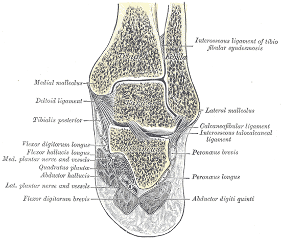
踝穴由胫骨穹窿、内踝、外踝(腓骨)与距骨构成;下胫腓联合(前后联合韧带、骨间韧带)维持踝穴稳定。The mortise comprises the tibial plafond, medial and lateral (fibular) malleoli and the talus; the syndesmosis keeps it stable.
机制:足的旋前/旋后 + 外旋/内收暴力;Weber 分型只看腓骨骨折线相对下胫腓联合的位置:A 在下、B 平齐、C 在上。Mechanism: pronation/supination + rotation/adduction forces; Weber looks only at the fibular fracture level vs the syndesmosis: A below, B at, C above.
位置越高,下胫腓联合损伤可能越重。The higher the level, the greater the potential syndesmotic injury.
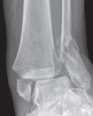
图 5-1 · 踝穴冠状切面(公有领域)Fig 5-1 · Mortise coronal section (public domain) 观察点:右距小腿关节与距跟关节冠状切面:胫骨延续为内踝、腓骨延续为外踝——两骨为独立骨块;下胫腓骨间韧带(联合)位于两骨之间;距骨坐于踝穴内、胫骨穹窿之下。这正是"踝穴由胫骨穹窿、内外踝与距骨构成、下胫腓联合维持稳定"的解剖对应。Observation: coronal section through the right talocrural and talocalcaneal joints: the tibia continues as the medial malleolus and the fibula as the lateral malleolus — two separate bones; the syndesmotic interosseous ligament lies between them; the talus sits in the mortise beneath the tibial plafond. This is the anatomic correlate of "the mortise comprises the tibial plafond, malleoli and talus, stabilized by the syndesmosis". 能证明:踝穴的正常骨性结构(两块独立骨 + 联合 + 距骨)。不能证明:骨折或联合损伤;1918 年解剖图谱,非影像。Shows: the normal bony architecture of the mortise (two separate bones + syndesmosis + talus). Does not show: fractures or syndesmotic injury; a 1918 anatomy plate, not imaging. 来源:Henry Gray《Anatomy of the Human Body》(1918) Plate 357,绘图 Henry Vandyke Carter,公有领域 · Wikimedia Commons 原页 · 独立图例Source: Henry Gray, Anatomy of the Human Body (1918), Plate 357, illustrated by Henry Vandyke Carter, public domain · Wikimedia Commons file page · standalone figure图 5-2 · 双踝骨折正位 X 线(CC BY 4.0)Fig 5-2 · Bimalleolar fracture frontal X-ray (CC BY 4.0) 观察点:踝关节正位 X 线:内踝骨折伴移位、外踝(腓骨远端)骨折,即双踝骨折。双踝骨折意味着踝穴环两处断裂,多不稳定,常需切开复位内固定以恢复踝穴——对应"不稳定 / 双踝 / 三踝:切开复位内固定"的治疗原则。Observation: frontal ankle X-ray: displaced medial malleolar fracture plus distal fibular (lateral malleolar) fracture — a bimalleolar fracture. Two breaks in the malleolar ring usually mean instability, often requiring ORIF to restore the mortise — matching the "unstable / bi- / trimalleolar: ORIF" principle. 能证明:双踝骨折的 X 线表现(内外踝均骨折)。不能证明:下胫腓联合是否损伤(需应力位/CT);与图 5-1 非同一来源。Shows: the X-ray appearance of bimalleolar fracture (both malleoli fractured). Does not show: whether the syndesmosis is injured (needs stress views/CT); not the same source as Fig 5-1. 来源:Tellisi N, Abdulkareem IS, Giannoudis PG,"Combined ipsilateral calcaneal and bimalleolar ankle fractures treated by open reduction and internal fixation",OA Case Reports 2012;1(1),DOI 10.13172/2052-0077-1-1-288,经 Wikimedia Commons 以 CC BY 4.0 发布(许可)· 原页 · 独立病例Source: Tellisi N, Abdulkareem IS, Giannoudis PG, "Combined ipsilateral calcaneal and bimalleolar ankle fractures treated by open reduction and internal fixation", OA Case Reports 2012;1(1), DOI 10.13172/2052-0077-1-1-288, via Wikimedia Commons under CC BY 4.0 (license) · file page · standalone case
初评和红旗Initial assessment & red flags
看 → 摸 → 动:畸形、肿胀、瘀斑;压痛部位;观察自然体位与自主活动受限,不主动诱发不稳。Look → feel → move: deformity, swelling, ecchymosis; tender points; resting position and limited spontaneous motion; do not deliberately provoke instability.
神经血管:足背动脉、足部感觉运动;初评及每次复位操作前后都要记录。Neurovascular: pedal pulses, foot sensation/motion; document at initial assessment and before and after each reduction.
红旗Red flags
开放骨折:踝部皮下就是骨头,开放伤常见 → 按开放骨折处理。Open fracture: bone is subcutaneous at the ankle, so open injuries are common → manage as open fracture.
明显移位/脱位:即使计划手术,也要尽快复位,减轻软组织与神经血管压迫。Marked displacement/dislocation: reduce ASAP even if surgery is planned, to relieve soft-tissue and neurovascular compression.
张力性水疱、皮肤发亮发白:软组织危象的信号。Tension blisters, shiny pale skin: signals of soft-tissue crisis.
糖尿病、血管病变:显著增加伤口并发症风险,需专科个体化评估、优化风险并制定稳定策略,而非自动更保守。Diabetes, vascular disease: significantly raise wound-complication risk; they need specialist individualized assessment, risk optimization and a stability-oriented plan — not automatic conservatism.
应力位(重力应力位):帮助判断 Weber B 的稳定性;手法应力须由专科判断,不要主动诱发不稳;CT:看后踝骨折块与关节面。Stress views (gravity stress): help judge stability in Weber B; manual stress needs specialist judgment — do not actively provoke instability; CT: assess the posterior malleolar fragment and joint surface.
分类 ≠ 手术指征Classification ≠ indication:Weber 告诉你骨折线在哪;做不做手术看的是踝穴稳不稳定(距骨移位、下胫腓联合)与软组织。: Weber tells you where the fibular line is; the surgical question is mortise stability (talar shift, syndesmosis) plus soft tissue.
AO 原则如何适用How AO principles apply
恢复腓骨长度与旋转:腓骨短了、转了,踝穴就不对。Restore fibular length and rotation: a short or rotated fibula means a malreduced mortise.
踝穴解剖复位:距骨必须正位于踝穴中央。Anatomic mortise reduction: the talus must sit centered.
稳定的下胫腓联合:联合不稳要固定(定位螺钉等方式,遵术式规范)。Stable syndesmosis: fix it when unstable (positioning screws etc., per technique standards).
固定顺序没有通用模板:按 CT 骨折形态、手术入路、软组织条件与稳定性要求制定;例如经后外侧入路可先固定后踝(Volkmann 块)+ 腓骨,再经内侧入路固定内踝(见 AO 官方页面)。There is no universal fixation sequence: plan the order from CT morphology, surgical approach, soft tissue and stability needs; e.g., via a posterolateral approach the posterior malleolus (Volkmann fragment) + fibula may be fixed first, then the medial side (see AO page).
治疗选择与限制Treatment options & limitations
稳定、无移位(如孤立外踝无距骨移位):可非手术(支具/石膏),但需随访防移位。Stable, undisplaced (e.g., isolated lateral malleolus without talar shift): nonoperative (brace/cast) with follow-up against displacement.
不稳定 / 双踝 / 三踝:切开复位内固定,恢复踝穴。Unstable / bi- / trimalleolar: ORIF to restore the mortise.
肿胀明显:先复位 + 石膏托 + 抬高,分期手术(皮纹征后,仍需综合全身、软组织与感染情况、由专科判断再做)。Marked swelling: reduce + backslab + elevation, staged surgery (after the wrinkle sign, the decision still needs overall, soft-tissue and infection assessment with specialist judgment).
限制:糖尿病/血管病变(伤口风险高,需个体化优化)、严重肿胀、依从性差——都可能让方案调整或分期。Limits: diabetes/vascular disease (high wound risk, needs individualized optimization), severe swelling, poor compliance — all may modify the plan or require staging.
Q1 · 22岁男性,扭伤后右踝肿痛,X线示腓骨骨折线在下胫腓联合水平(Weber B),内踝压痛明显。查体还必须做哪一项?(合成教学病例)Q1 · A 22-year-old man twisted his ankle; X-ray shows a fibular fracture at the syndesmosis (Weber B) with marked medial tenderness. What exam step is still mandatory? (synthetic teaching case)
回答正确 ✓Correct ✓
再想想 ✗Try again ✗
答案:C。Answer: C.任何踝骨折都要摸全长腓骨:近端压痛提示 Maisonneuve 损伤,漏诊会导致下胫腓不稳被忽视。Always palpate the full fibula: proximal tenderness suggests Maisonneuve injury; missing it leaves syndesmotic instability unaddressed.
Q2 · Weber 分型的依据是?Q2 · The Weber classification is based on?
回答正确 ✓Correct ✓
再想想 ✗Try again ✗
答案:B。Answer: B.Weber 只看一点:腓骨骨折线在联合水平下(A)、平齐(B)还是上(C)。记住:分类描述形态,不直接等于手术指征。Weber asks one thing: the fibular line below (A), at (B), or above (C) the syndesmosis. Remember: classification describes morphology; it does not equal indication.
AO Trauma — ORP Handout: Malleolar fractures (PDF, official) 定位:官方教学讲义;注意固定顺序无通用模板,须按骨折形态、入路与软组织制定(见新增 AO 页面)。访问日期 2026-10-04。Relevance: official teaching handout; note there is no universal fixation sequence — plan it from fracture morphology, approach and soft tissue (see new AO page). Accessed 2026-10-04.
AO Surgery Reference — ORIF for transsyndesmotic, posterior, lateral simple, and medial fractures 定位:固定顺序按情况制定——经后外侧入路可先固定后踝(Volkmann 块)+ 腓骨、再固定内侧;或先内外侧、后以前方螺钉固定后踝;内外侧孰先由术者判断。访问日期 2026-10-04。Relevance: fixation order is case-dependent — via a posterolateral approach the posterior malleolus (Volkmann fragment) + fibula may be fixed first, then the medial side; or medial/lateral first with anterior screws for the posterior fragment; medial-vs-lateral priority is the surgeon's judgment. Accessed 2026-10-04.
媒体来源:图 5-1 为公有领域解剖图(Gray's Anatomy 1918);图 5-2 CC BY 4.0(Tellisi N 等,《OA Case Reports》2012),随包附带、原页与许可见图注;AO Surgery Reference / AO Trauma 为外部来源链接,仅引用未复制其图文与视频,内容版权归 AO Foundation 所有(AO 未提供公开再发布许可)。Media sources: Fig 5-1 is a public-domain anatomy plate (Gray's Anatomy, 1918); Fig 5-2 CC BY 4.0 (Tellisi N et al., OA Case Reports 2012), bundled with source page and license in its caption; AO Surgery Reference / AO Trauma are external sources linked for reference only — no content copied; copyright remains with the AO Foundation (AO offers no open redistribution license).
临床判断与关键步骤Clinical decisions and key steps
具体方案;以本院为准;每项数字同时核对人群、阶段和来源。Follow institutional protocols; check population, stage and source with every number.
开放骨折:抗生素、清创与覆盖Open fracture: antibiotics, debridement and coverage
抗生素依据并列Antibiotic sources side by side
尽早静脉抗生素,尽量伤后1 h内。国内2019 Gustilo I–II以一代头孢(原文头孢唑啉)为基础;III加氨基糖苷,粪便/厌氧污染加青霉素或甲硝唑;过敏/肾功能及本地耐药谱决定替代。以本院规范为准。Early IV antibiotics, ideally within one hour. Chinese 2019 GustiloI–II first-generation cephalosporin(cefazolin), III adds aminoglycoside, fecal/anaerobic contamination adds penicillin/metronidazole; allergy/renal function/local resistance guide alternatives. Follow institutional policy.
国内2019持续至闭合后48 h;不能当所有现代方案统一疗程,按本院抗菌管理选择。盐水湿敷+封闭层与授权照片;BOAST/NICE不做急诊小冲洗,国内旧文急诊清洗说法另需本院定标。以本院规范为准。Chinese 2019 continues until48 hours after closure, not a universal modern duration; use local stewardship. Saline-moist/occlusive dressing and authorized photo; BOAST/NICE no ED mini-washout differs from older Chinese cleaning text. Follow institutional policy.
清创与重建Debridement and reconstruction
重污染/血管受损立即;其他高能12 h内、低能24 h内清创;确定覆盖尽量72 h内,内固定需与覆盖配合。破伤风与多区域复评同步。以本院规范为准。Immediate debridement for severe contamination/vascular compromise; other high-energy within 12 hours/low-energy within 24; definitive coverage ideally within 72, coordinated with fixation. Include tetanus and whole-patient review. Follow institutional policy.
筋膜室:早诊与立即减压Compartment syndrome: early diagnosis and immediate decompression
有脉搏也可缺血A pulse can coexist with ischemia
高风险每小时查体,记录疼痛/被动牵拉痛及镇痛剂量;有症状松解环形层至皮肤,心脏水平,30 min内复评并即时骨科会诊。以本院规范为准。High risk: hourly exam, pain/passive stretch/analgesic dose. Symptoms: release circumferential layers to skin, heart-level limb, reassess within 30 minutes and consult orthopedics immediately. Follow institutional policy.
DBP−间室压<30 mmHg风险增加;绝对压>40 mmHg考虑紧急减压,不能脱离临床。确诊后立即切开;国内2019“6–8 h内”是旧表述,不能作为等待理由。以本院规范为准。DBP minus compartment pressure below 30 increases risk; absolute over 40 prompts urgent decompression assessment in context. Once diagnosed, immediate fasciotomy; older Chinese 2019 wording within 6–8 hours is not permission to delay. Follow institutional policy.
肢体血管伤:没有安全等待时间Limb vascular injury: no safe waiting time
硬征与紧急复流Hard signs and urgent reperfusion
无脉、持续出血、扩张性血肿为硬征;毛细血管再充盈或Doppler不能单独排除。立即骨科/血管/整形协同,必要时临时分流。以本院规范为准。Absent pulse/ongoing bleeding/expanding hematoma are hard signs; CRT/Doppler alone cannot exclude injury. Immediate orthopedic/vascular/plastic coordination, temporary shunt when needed. Follow institutional policy.
BOAST2026:到院1 h内开始血运重建(commenced),不是承诺1 h内已恢复;不因复杂影像延误复流。以本院规范为准。BOAST 2026: commence revascularization within one hour of arrival, not a guarantee of completed flow restoration by then; complex imaging must not delay reperfusion. Follow institutional policy.
老年髋部:早镇痛、早MDT、限期手术Geriatric hip: analgesia, MDT and timely surgery
时点与条件Timing and conditions
国内2022:完善术前准备后力争入院48 h内完成;NICE入院当天或次日。两者均需及时纠正可治疗合并症,不能以48 h为等待目标。以本院规范为准。Chinese 2022: aim to complete within 48 hours after admission with preparation; NICE same/next day. Correct treatable comorbidity promptly;48 hours is not a target to wait for. Follow institutional policy.
神经阻滞可由受训者用于镇痛不足或限制阿片;髂筋膜阻滞“国内首选”尚未核对附件原句,暂不宣称;不能替代早手术。以本院规范为准。Trained nerve block may supplement inadequate analgesia/limit opioids. The national first-choice fascia-iliaca wording is not confirmed from the attachment; no such claim is made. Blocks do not replace early surgery. Follow institutional policy.
本区重点:查体、血供与升级Regional focus: examination, perfusion and escalation
下肢:局部与全身风险Lower limb: local and systemic risk
膝脱位(包括自行复位):复位前后查血管与腓总神经,测ABI;<0.9或脉搏不对称行CTA/血管外科评估。≥0.9仍需序贯查体,不能只因有脉搏排除(BASK)。Knee dislocation, including spontaneously reduced injury: assess vessels/peroneal nerve and ABI before/after reduction. Below 0.9 or asymmetric pulses requires CTA/vascular review. Serial examinations remain necessary at ≥0.9 (BASK).
股骨干牵引夹板仅用于适合的孤立股骨干伤;合并骨盆、同侧膝/小腿/踝损伤避免牵引,先选其他固定并核对装置(院前共识/技能规范)。Traction splints suit selected isolated femoral shaft injuries; avoid traction with pelvic or ipsilateral knee/lower-leg/ankle injury. Use alternative stabilization and verify device rules (prehospital guidance).
胫骨骨折为筋膜室高风险:序贯疼痛、牵拉痛、感觉/运动及镇痛量;疑诊立即松解束缚与骨科评估,脉搏存在不排除(BOAST)。Tibial fractures carry compartment risk: serial pain/stretch, sensory/motor and analgesic-dose assessment. Release constriction and obtain urgent orthopedic review when suspected; pulses do not exclude it (BOAST).
老年髋部骨折走老年共管路径;NICE入院当天或次日手术,国内48 h目标需完成必要优化,神经阻滞不替代早期手术(NICE/国内实施依据)。Older hip-fracture patients need a geriatric co-care pathway: NICE surgery on admission day/next day; the Chinese 48-hour goal requires necessary optimization. Nerve blocks do not replace early surgery (NICE/Chinese implementation evidence).