成人创伤教学 · 基于 AO Surgery Reference 官方原则 · 配图为真实影像(公开许可)Adult trauma teaching · Based on official AO Surgery Reference principles · Illustrated with real, openly licensed images
预计学习Estimated learning time · 60 分钟min
学完这门课,你能……After this course, you can…
描述骨折、脱位与软组织风险。Describe fracture, dislocation and soft-tissue risk.
核对远端感觉、运动和灌注。Assess distal sensation, motor function and perfusion.
比较处理前后变化并组织复评。Compare findings around interventions and organize reassessment.
实习:识别与报告;规培:决策时机与监督下关键步骤;青年主治:团队指挥与资源调度。Intern: recognize/report; resident: decision timing and supervised key steps; early attending: team leadership and resources.
学习说明How to use 每单元按统一框架组织:解剖与机制 → 初评和红旗 → 影像/分类 → AO 原则如何适用 → 治疗选择与限制 → 并发症/随访 → 病例自测。正文为短句,细节放在折叠区;病例支持分段揭晓。每单元末附来源与媒体许可。Each unit follows the same framework: Anatomy & Mechanism → Initial Assessment & Red Flags → Imaging / Classification → How AO Principles Apply → Treatment Options & Limits → Complications / Follow-up → Case Self-Test. Main text uses short sentences; details sit in collapsible sections; cases support step-by-step reveal. Sources and media licences close each unit.
重要原则Key principle 分类描述损伤形态,不等于手术指征。治疗决策需结合患者年龄、功能需求、骨质量、合并伤与医疗条件综合判断。本课程为成人教学,不涉及儿童骨折。Classification describes the injury pattern — it is not, by itself, an indication for surgery. Treatment decisions must integrate the patient's age, functional demands, bone quality, associated injuries and available resources. This course covers adult trauma only.
关于图像About the images 本课程配图为真实 X 线/CT(Wikimedia Commons 与 PMC 开放获取,CC0/CC BY/CC BY-SA,逐图核验许可并随包附原文件,清单见 assets/media_sources.csv)。每张图为独立病例图片,不同图片非同一患者,不构成连续病例序列;图注说明实际所见与局限。AO Surgery Reference 图例仅以外链引用原官方页面,未复制、未热链其图片。All figures in this course are real radiographs/CT images (Wikimedia Commons and PMC open access, CC0/CC BY/CC BY-SA; each licence verified and originals bundled — see assets/media_sources.csv). Each image is from an independent case; different images are not the same patient and do not form a case sequence. Captions describe what is actually seen and the limitations. AO Surgery Reference illustrations are referenced by link to the official pages only — no images copied or hotlinked.
1肩关节脱位:单纯脱位 vs 骨折脱位Shoulder Dislocation: Simple vs Fracture-Dislocation
1.1 解剖与机制1.1 Anatomy & Mechanism
盂肱关节是典型的球窝关节,活动度大,稳定性主要依赖软组织:盂唇、关节囊、肩袖与韧带。The glenohumeral joint is a classic ball-and-socket joint with a large range of motion; stability relies mainly on soft tissues — labrum, capsule, rotator cuff and ligaments.
前脱位最常见。典型机制:上肢外展外旋位受暴力,或跌倒时手撑地。Anterior dislocation is the most common. Typical mechanism: force applied with the arm abducted and externally rotated, or a fall onto the outstretched hand.
后脱位少见,多见于电击伤、癫痫发作或高能量创伤,容易漏诊。Posterior dislocation is uncommon, seen after electric shock, seizures or high-energy trauma, and is easily missed.
单纯脱位:仅关节脱位,无骨折。骨折脱位:脱位合并肱骨近端骨折,最常见为大结节骨折。Simple dislocation: dislocation without fracture. Fracture-dislocation: dislocation with a proximal humerus fracture, most commonly of the greater tuberosity.
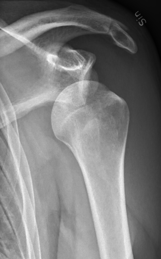
图 1-1 Fig 1-1 真实 X 线:左肩正位(复位前),肱骨头向前下移位、关节盂空虚,为典型的前下脱位。局限:单张正位片仅展示一种脱位方向;实际阅片需结合穿胸位/Y 位等多体位判断方向并排除合并骨折。独立来源影像,与练习病例无关。Real radiograph: AP left shoulder before reduction; the humeral head is displaced anterior-inferior with an empty glenoid — a typical anterior-inferior dislocation. Limitation: a single AP view shows one direction only; real interpretation needs additional projections to confirm direction and exclude associated fracture. Independent source image, separate from the practice vignette.图片:Mikael Häggström, M.D.(Wikimedia Commons),CC0 1.0 公有领域贡献(原文件页 · 许可)Image: Mikael Häggström, M.D. (Wikimedia Commons), CC0 1.0 public-domain dedication (file page · licence)展开:为什么前脱位最常见?Expand: why is anterior dislocation the most common?
关节盂浅、前下方骨性约束最弱,外展外旋时肱骨头容易向前下方滑出。The glenoid is shallow and bony constraint is weakest anteroinferiorly; in abduction-external rotation the humeral head slides out anteroinferiorly.
前脱位常合并 Bankart 损伤(前下盂唇撕脱)与 Hill-Sachs 损伤(肱骨头后外侧压缩骨折),是复发性不稳定的解剖基础。Anterior dislocation is often accompanied by a Bankart lesion (anteroinferior labral detachment) and a Hill-Sachs lesion (posterolateral humeral head compression fracture) — the anatomic basis of recurrent instability.
1.2 初评和红旗1.2 Initial Assessment & Red Flags
先看全身:多发伤患者按 ATLS 流程,肩部不是优先处理部位。Check the whole patient first: in polytrauma follow ATLS; the shoulder is not the first priority.
视诊:方肩畸形、患肢轻度外展弹性固定。Inspection: squared-off shoulder contour, arm held in slight abduction with elastic resistance.
神经血管检查必须在复位前、后各做一次并记录。Neurovascular examination is mandatory before and after reduction, and must be documented.
红旗Red flags
腋神经损伤:三角肌区(上臂外侧)感觉减退、主动外展无力。Axillary nerve injury: reduced sensation over the deltoid region (lateral upper arm), weak active abduction.
血管损伤体征:桡动脉搏动减弱/消失、手部苍白发凉(少见但危重)。Vascular injury signs: diminished/absent radial pulse, pale cool hand (rare but critical).
皮肤破损/开放性脱位:按开放性损伤流程处理。Skin breach / open dislocation: manage as an open injury.
复位后仍有神经症状或血管体征:立即上级评估。Persistent neurologic symptoms or vascular signs after reduction: escalate immediately.
1.3 影像 / 分类1.3 Imaging / Classification
常规:肩关节正位 + 腋位(或 Y 位),确认脱位方向、排除骨折。Routine: AP + axillary (or Y) views to confirm direction and exclude fracture.
怀疑骨折脱位、骨缺损或复位困难:加做 CT(含三维重建)。If fracture-dislocation, bony defect or difficult reduction is suspected: add CT (with 3D reconstruction).
复位后必须复查 X 线,确认肱骨头同心圆复位。Post-reduction radiographs are mandatory to confirm concentric reduction of the humeral head.
展开:分类(方向 × 是否合并骨折)Expand: classification (direction × fracture or not)
按方向:前脱位、后脱位、下脱位(盂下)、上脱位(罕见)。By direction: anterior, posterior, inferior (luxatio erecta), superior (rare).
分类用于描述损伤,不等于手术指征。是否手术取决于骨折移位、关节稳定性与患者因素。Classification describes the injury — it is not an indication for surgery. Operative decisions depend on fracture displacement, joint stability and patient factors.
1.4 AO 原则如何适用1.4 How AO Principles Apply
AO 四原则:解剖复位、稳定固定、保护血供、早期功能锻炼——主要针对骨折;且“解剖复位”按部位理解:关节面追求解剖复位,骨干恢复长度、对线与旋转即可。The four AO principles — anatomic reduction, stable fixation, preservation of blood supply, early functional exercise — apply primarily to fractures; and "anatomic reduction" is site-specific: articular surfaces pursue anatomic reduction, while diaphyseal fractures need length, alignment and rotation restored.
稳定性与之匹配:关节内骨折多需绝对稳定,骨干骨折相对稳定下的二期愈合同样是 AO 认可的路径。Match stability to the site: intra-articular fractures usually need absolute stability; diaphyseal fractures heal well by secondary intention under relative stability — equally an AO-recognized pathway.
单纯脱位:核心是恢复关节同心圆对合(复位),然后通过短期制动 + 渐进康复恢复功能。Simple dislocation: the core is restoring concentric joint congruity (reduction), then short immobilisation plus progressive rehabilitation.
骨折脱位:按骨折处理——先评估骨折块,再决定复位顺序与固定方式;保护肱骨头血供是关键考量。Fracture-dislocation: manage as a fracture — assess fragments first, then decide the reduction sequence and fixation; protecting humeral head blood supply is a key consideration.
早期功能锻炼适用于稳定复位之后;不稳定的关节过早活动会导致再脱位。Early mobilisation applies after a stable reduction; moving an unstable joint too early risks re-dislocation.
1.5 治疗选择与限制1.5 Treatment Options & Limits
单纯脱位Simple闭合复位为主Closed reduction first
在充分镇静镇痛下轻柔复位;忌暴力。Reduce gently under adequate sedation and analgesia; never use force.
复位后:复查神经血管 + X 线;悬吊短期制动,制动时长个体化。After reduction: recheck neurovascular status + radiographs; sling immobilisation for a short, individualised period.
年轻、运动需求高的首发前脱位患者复发风险高,需康复随访与复发预案。Young, active patients with a first-time anterior dislocation carry a high recurrence risk — arrange rehabilitation follow-up and a recurrence plan.
骨折脱位Fracture-dislocation按骨折原则处理Managed by fracture principles
先明确骨折块大小、移位与关节面受累(CT 与否依据骨折复杂度和临床稳定性决定),再定方案;若合并血管损伤等危及肢体的情况,紧急处置优先,不因等待影像而延误。First define fragment size, displacement and articular involvement (CT as fracture complexity and clinical stability dictate), then decide; with vascular injury or other limb-threatening conditions, urgent management takes priority and must not wait for imaging.
大结节明显移位、关节面劈裂或复位后不稳定:多需手术(固定或按肱骨近端骨折处理)。Markedly displaced greater tuberosity, articular head-split or post-reduction instability: usually operative (fixation, managed as a proximal humerus fracture).
无明显移位的骨折脱位:可在麻醉下复位后按肱骨近端骨折保守治疗并密切随访。Minimally displaced fracture-dislocation: may be reduced under anaesthesia and managed non-operatively as a proximal humerus fracture with close follow-up.
展开:治疗的限制与注意事项Expand: limits and cautions
本课程不规定具体复位手法与药物剂量;手法选择与镇静方案遵循本院流程与麻醉科协作。This course does not prescribe specific reduction manoeuvres or drug doses; follow local protocols with anaesthesia support.
骨质疏松患者:骨折脱位风险更高,固定选择需考虑骨质量。Osteoporotic patients: higher fracture-dislocation risk; fixation choices must account for bone quality.
陈旧性脱位(数周未复位):不要强行闭合复位,需专科评估。Chronic dislocation (unreduced for weeks): do not force closed reduction — refer for specialist assessment.
1.6 并发症 / 随访1.6 Complications / Follow-up
复发性脱位 / 慢性不稳定:尤其年轻首发前脱位患者。Recurrent dislocation / chronic instability: especially young patients after a first anterior dislocation.
骨缺损相关不稳:Bankart 与 Hill-Sachs 病变进展。Bony-defect-related instability: progression of Bankart and Hill-Sachs lesions.
腋神经损伤:多数为牵拉伤可恢复,需随访肌力与感觉。Axillary nerve injury: usually a traction injury that recovers; follow strength and sensation.
创伤性关节炎:反复脱位或关节面损伤后远期风险。Post-traumatic arthritis: long-term risk after recurrent dislocation or articular injury.
注:本病例为合成教学病例,非真实患者。Note: synthetic teaching case, not a real patient.
28 岁男性,篮球争抢篮板后右肩剧痛,不敢活动。点击按钮逐步揭晓诊疗思路。A 28-year-old man has severe right shoulder pain and refuses to move it after contesting a rebound. Reveal the workup step by step.
步骤 1 · 病史与查体Step 1 · History & exam 方肩畸形,患肢弹性固定于轻度外展位。桡动脉搏动可及,手部温暖。三角肌区感觉减退,主动外展不能(疼痛限制 vs 神经损伤,待鉴别)。Squared-off shoulder; arm elastically fixed in slight abduction. Radial pulse palpable, hand warm. Reduced sensation over the deltoid; active abduction impossible (pain vs nerve injury — to be clarified).
步骤 2 · 影像Step 2 · Imaging 正位 + 腋位 X 线:肱骨头位于关节盂前下方,未见明确骨折线。诊断:单纯性肩关节前脱位。AP + axillary radiographs: humeral head anteroinferior to the glenoid, no definite fracture line. Diagnosis: simple anterior shoulder dislocation.
步骤 4 · 后续Step 4 · Follow-up 悬吊短期制动后转康复:活动度→力量→本体感觉。告知年轻首发前脱位复发风险高,若再脱位或出现交锁感及时复诊。Short sling immobilisation then rehabilitation: motion → strength → proprioception. Counsel that first-time anterior dislocation in a young patient carries high recurrence risk; return promptly if it re-dislocates or locking occurs.
Q1. 肩关节前脱位复位后,最关键的即刻复查是什么?Q1. What is the most important immediate recheck after reducing an anterior shoulder dislocation?
答案:CAnswer: C 复位前后都必须做神经血管检查并记录,X 线确认肱骨头真正复位(排除骨折脱位漏诊)。过早全范围活动(A)可致再脱位;D 无指征。Neurovascular status must be checked and documented before and after reduction, and radiographs must confirm true concentric reduction (excluding a missed fracture-dislocation). Early full motion (A) risks re-dislocation; (D) is not indicated.
Q2. 同一患者 X 线若同时显示大结节骨折块明显移位,处理策略应如何调整?Q2. If the same patient's radiograph also shows a markedly displaced greater tuberosity fragment, how should management change?
答案:BAnswer: B 骨折脱位 ≠ 单纯脱位。应先明确骨折块情况(CT 与否依据骨折复杂度和临床稳定性决定),明显移位的大结节骨折多需手术固定;暴力复位(D)可能加重骨折移位或损伤血供。Fracture-dislocation ≠ simple dislocation. Define the fracture pattern first (CT as complexity and stability dictate); a markedly displaced greater tuberosity usually needs operative fixation. Forceful reduction (D) may worsen displacement or injure blood supply.
对照官方图例,复习损伤识别与处理原则:Review injury recognition and management principles alongside the official illustrations:
AO Surgery Reference — 肱骨近端骨折脱位定义 肱骨近端骨折脱位的 AO 定义与分类框架;单纯肩脱位(无骨折)AO 无独立页面AO definition and classification framework for proximal humerus fracture-dislocation; AO has no standalone simple-dislocation page
AAOS OrthoInfo — 肩关节脱位 单纯肩脱位:解剖、前脱位最常见、查体与影像、急诊镇静下轻柔复位Simple shoulder dislocation: anatomy, anterior most common, examination and imaging, gentle reduction under sedation in the emergency setting
注:依 AO FAQ,AO 图例仅允许非商业演示使用;在网站转载需申请许可。本课程仅外链官方原页面,未复制、未热链任何 AO 图片。Note: per the AO FAQ, AO illustrations may be used in non-commercial presentations only; republication on a website needs permission. This course links to the official pages and copies or hotlinks no AO images.
本单元来源与媒体许可Sources & media licence — Unit 1
注:“来源(已核验)”仅指该网页于访问日期可正常打开、标题与定位描述相符;不构成对本课程每条医学表述的逐条医学审核。临床决策请结合最新指南与本院流程。Note: "Source (verified)" means only that the page was accessible on the access date and that its title and scope description match; it does not constitute item-by-item medical review of every statement in this course. Base clinical decisions on current guidelines and local protocols.
来源 1(已核验)Source 1 (verified) Extraarticular 3-part, surgical neck and tuberosity, dislocation — definition 定位:肱骨近端骨折脱位定义页(AO Surgery Reference,成人创伤模块);支撑本单元“骨折脱位”部分的分类与 AO 原则适用性。Scope: proximal humerus fracture-dislocation definition page (AO Surgery Reference, adult trauma); supports the fracture-dislocation classification and AO-principles sections of this unit. 访问日期:2026-10-04Accessed: 2026-10-04
来源 2(已核验 · AAOS,非 AO)Source 2 (verified · AAOS, not AO) Shoulder Dislocation - OrthoInfo - AAOS 定位:美国骨科医师学会(AAOS)OrthoInfo 患者教育页面(由美国肩肘外科医师学会 ASES 成员撰写/审核);涵盖肩关节解剖、前脱位最常见、电击/癫痫致后脱位、查体(神经血管)、影像(X 线确诊方向与骨折,CT/MRI 指征)与治疗(急诊镇静下轻柔闭合复位、悬吊制动、康复、手术指征);支撑本单元“单纯脱位”章节。明确标注:AAOS 来源,非 AO Surgery Reference。Scope: American Academy of Orthopaedic Surgeons (AAOS) OrthoInfo patient-education page (written/reviewed by an American Shoulder and Elbow Surgeons member); covers shoulder anatomy, anterior dislocation most common, seizure/electric-shock posterior dislocation, examination (neurovascular), imaging (radiographs confirm direction and fracture; CT/MRI indications) and treatment (gentle closed reduction under sedation in the emergency setting, sling immobilisation, rehabilitation, surgical indications); supports this unit's "simple dislocation" sections. Clearly labelled: AAOS source, not AO Surgery Reference. 访问日期:2026-10-04Accessed: 2026-10-04
诚实标注:AO Surgery Reference 以骨折治疗为核心,未在其官方域名内找到“单纯肩关节脱位(无骨折)独立处理”页面;本单元“单纯脱位”章节改引 AAOS OrthoInfo 来源(来源 2,患者教育页面)。不可将 AO 骨折脱位页面的表述外推为单纯脱位的处理依据。Honesty note: AO Surgery Reference is fracture-treatment oriented; no standalone simple-shoulder-dislocation (no fracture) page exists on its domain, so this unit's "simple dislocation" sections cite the AAOS OrthoInfo source (Source 2, patient-education page) instead. Do not extrapolate AO fracture-dislocation statements to simple-dislocation management.
本单元图片(图 1-1):真实 X 线/CT,逐图核验许可;作者、原标题、原 URL、许可与原 caption 见 assets/media_sources.csv,各图注含原文件页与许可链接。各图为不同患者的独立图片,非同一病例序列。Unit images (Fig 1-1): real radiographs/CT, licences verified per image; author, original title, source URL, licence and original caption are listed in assets/media_sources.csv, and each caption links its file page and licence. Images are from different patients, not a case sequence.
2肘关节脱位:单纯脱位 vs 骨折脱位Elbow Dislocation: Simple vs Fracture-Dislocation
2.1 解剖与机制2.1 Anatomy & Mechanism
肘关节由肱尺、肱桡、上尺桡三个关节组成,稳定性依赖骨性结构(冠状突、鹰嘴)与内外侧副韧带。The elbow comprises the ulnohumeral, radiocapitellar and proximal radioulnar joints; stability depends on bony structures (coronoid, olecranon) and the collateral ligaments.
后脱位最常见,典型机制是跌倒时手撑地、肘过伸伴旋转暴力。Posterior dislocation is the most common; typical mechanism is a fall onto the outstretched hand with hyperextension and rotational force.
单纯脱位:无骨折。骨折脱位(复杂脱位):合并桡骨头 / 冠状突 / 鹰嘴等骨折。Simple dislocation: no fracture. Fracture-dislocation (complex dislocation): with fractures of the radial head, coronoid, olecranon, etc.
恐怖三联征 = 肘关节后脱位 + 桡骨头骨折 + 冠状突骨折,常合并外侧副韧带损伤,高度不稳定。The terrible triad = posterior elbow dislocation + radial head fracture + coronoid fracture, often with lateral collateral ligament injury — highly unstable.
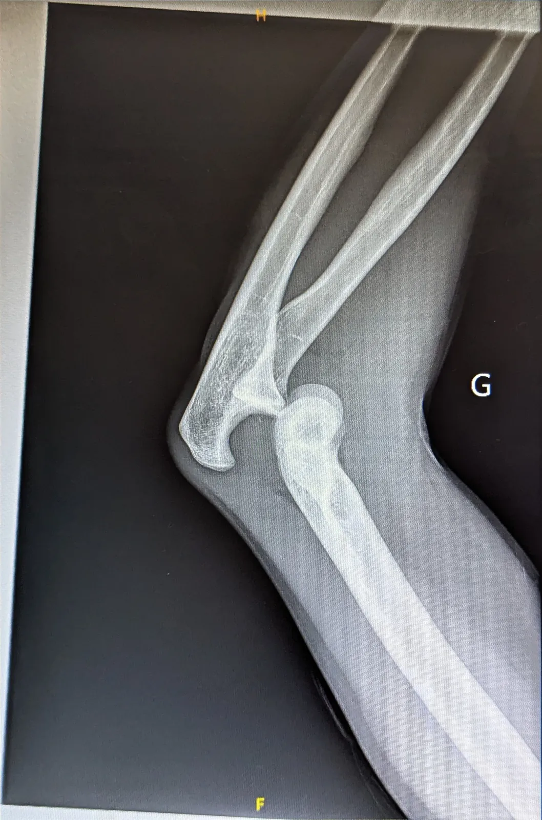
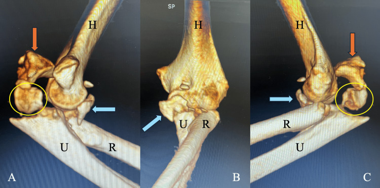
图 2-1 Fig 2-1 真实 X 线:左肘侧位,尺骨近端/鹰嘴向肱骨远端后方移位,为肘关节后脱位,未见明显骨折线。局限:本图为翻拍 X 线胶片的照片,细节较直接数字片偏软;单张侧位片不能排除隐匿骨折,需结合正位片与查体。独立来源影像,与练习病例无关。Real radiograph: lateral left elbow; the proximal ulna/olecranon is displaced posterior to the distal humerus — a posterior elbow dislocation with no obvious fracture line. Limitation: this is a photograph of an X-ray film, softer than a direct digital image; a single lateral view cannot exclude occult fracture — correlate with AP views and examination. Independent source image, separate from the practice vignette.图片:Flibust1er(Wikimedia Commons),CC0 1.0 公有领域贡献(原文件页 · 许可)Image: Flibust1er (Wikimedia Commons), CC0 1.0 public-domain dedication (file page · licence)图 2-2 Fig 2-2 真实 3D CT(标注为原作者所加):恐怖三联征的变异病例——后脱位(已复位)合并桡骨头骨折(黄圈)、冠状突骨折(蓝箭头),同时合并尺骨鹰嘴骨折(橙箭头)。局限:此为合并鹰嘴骨折的变异病例,非标准恐怖三联征;分辨率较低,仅作结构示意。独立来源影像,与练习病例无关。Real 3D CT (annotations by the original authors): a terrible-triad variant — posterior dislocation (reduced) with radial head fracture (yellow circle) and coronoid fracture (blue arrow), plus an associated olecranon fracture (orange arrow). Limitation: this is a variant case with an olecranon fracture, not a classic terrible triad; low resolution, for structural illustration only. Independent source image, separate from the practice vignette.图片:Sachinis 等,Cureus 病例报告 Figure 1(PMC12999191),CC BY 4.0(原文 · 许可)Image: Sachinis et al., Cureus case report, Figure 1 (PMC12999191), CC BY 4.0 (article · licence)展开:为什么冠状突如此重要?Expand: why is the coronoid so important?
冠状突是肘关节前方的骨性阻挡,缺损越大,肘关节越容易向后半脱位。The coronoid is the anterior bony buttress of the elbow; the larger the defect, the more prone the elbow is to posterior subluxation.
因此骨折脱位中冠状突骨折块的大小与移位,是判断稳定性的核心依据之一。Hence, in fracture-dislocation, the size and displacement of the coronoid fragment are core determinants of stability.
2.2 初评和红旗2.2 Initial Assessment & Red Flags
肘部畸形常很明显,但仍需按流程完成全身评估。Elbow deformity is often obvious, but the full systematic assessment still comes first.
复位前、后各做一次神经血管检查并记录:桡动脉搏动、正中/尺/桡神经功能。Perform and document a neurovascular examination before and after reduction: radial pulse and median/ulnar/radial nerve function.
红旗Red flags
肱动脉损伤:尤其合并肱骨远端/近端骨折时;搏动减弱、手部苍白发凉。Brachial artery injury: especially with associated distal/proximal humeral fractures; weak pulse, pale cool hand.
骨筋膜室综合征:前臂剧痛、被动牵拉痛、张力性肿胀。Compartment syndrome: severe forearm pain, pain on passive stretch, tense swelling.
开放性脱位 / 皮肤破损:按开放性损伤处理。Open dislocation / skin breach: manage as an open injury.
复位后仍不稳定、反复半脱位:提示关键稳定结构损伤,需进一步评估。Persistent instability or recurrent subluxation after reduction: suggests injury to key stabilisers — further evaluation needed.
2.3 影像 / 分类2.3 Imaging / Classification
常规:肘关节正侧位 X 线,判断脱位方向、有无骨折。Routine: AP and lateral elbow radiographs to determine direction and detect fractures.
怀疑骨折脱位:CT 明确桡骨头、冠状突骨折块的大小与移位。Suspected fracture-dislocation: CT to define the size and displacement of radial head and coronoid fragments.
复位后复查 X 线,确认关节同心圆复位、无残留半脱位。Post-reduction radiographs to confirm concentric reduction without residual subluxation.
按是否合并骨折:单纯脱位 vs 骨折脱位(含恐怖三联征)。By associated fracture: simple dislocation vs fracture-dislocation (including the terrible triad).
分类描述损伤,不等于手术指征;是否手术看复位后稳定性与骨折情况。Classification describes the injury, not the indication for surgery; operative decisions rest on post-reduction stability and the fracture pattern.
2.4 AO 原则如何适用2.4 How AO Principles Apply
肘关节的核心目标是:稳定 + 同心圆复位 + 早期活动。The core goals at the elbow are: stability + concentric reduction + early motion.
稳定固定:使关节在早期活动中保持同心圆对合,避免长期制动导致的僵硬。Stable fixation: keeps the joint concentrically reduced during early motion, avoiding stiffness from prolonged immobilisation.
保护血供与软组织:骨折脱位周围软组织损伤重,手术入路需保护重要结构。Preserve blood supply and soft tissues: soft-tissue injury is severe in fracture-dislocation; approaches must protect key structures.
2.5 治疗选择与限制2.5 Treatment Options & Limits
单纯脱位·稳定Simple · stable复位后早期活动Early motion after reduction
肘关节脱位属急症:尽快接受专业评估,在安全条件下复位(充分镇痛镇静、轻柔手法),复位前后检查神经血管与稳定性。Elbow dislocation is an emergency: seek prompt professional assessment and reduce under safe conditions (adequate sedation/analgesia, gentle manoeuvre), checking neurovascular status and stability before and after reduction.
复位后稳定:短期支具保护下尽早开始主动活动,避免长期制动。If stable after reduction: begin active motion as early as possible with short-term brace protection; avoid prolonged immobilisation.
骨折脱位Fracture-dislocation按损伤逐一处理,不套用同一方案Address each injury; no one-size-fits-all
桡骨头骨折:小块稳定可保守;移位明显或影响稳定 → 固定或置换(按骨折块大小与粉碎程度个体化)。Radial head fracture: small stable fragments may be non-operative; displaced or stability-compromising fractures → fixation or replacement, individualised to fragment size and comminution.
冠状突骨折:大块移位影响稳定 → 固定恢复前方骨性阻挡。Coronoid fracture: large displaced fragments compromising stability → fixation to restore the anterior buttress.
韧带损伤:外侧副韧带撕裂常需修复,以恢复旋转稳定性。Ligament injury: lateral collateral ligament tears often need repair to restore rotatory stability.
恐怖三联征:三个稳定结构均受损,多数需手术系统重建。Terrible triad: all three stabilisers injured; most need systematic operative reconstruction.
展开:限制与注意事项Expand: limits and cautions
肘关节最怕僵硬:能早期活动就不长期制动,能稳定固定就不靠石膏硬扛。The elbow's greatest enemy is stiffness: mobilise early when possible; don't rely on prolonged casting when stable fixation is achievable.
复位困难或复位后反复不稳:不要反复暴力尝试,及时请专科会诊。Difficult reduction or recurrent instability: don't repeat forceful attempts — get specialist help.
本课程不规定具体手术入路与内固定型号选择,遵循 AO Surgery Reference 与本院流程。This course does not prescribe specific approaches or implant choices; follow AO Surgery Reference and local protocols.
2.6 并发症 / 随访2.6 Complications / Follow-up
关节僵硬:最常见,康复是治疗的一部分。Joint stiffness: the most common; rehabilitation is part of treatment.
异位骨化:高能量损伤与手术后可能出现,影响活动度。Heterotopic ossification: may follow high-energy injury or surgery and limit motion.
尺神经症状、创伤性关节炎:中远期随访观察。Ulnar nerve symptoms, post-traumatic arthritis: watch for in mid- to long-term follow-up.
随访要点:活动度进展、稳定性复查、神经症状变化。Follow-up: progress of motion, stability recheck, changes in neurologic symptoms.
2.7 病例自测2.7 Case Self-Test
病例 2-1(分段揭晓)Case 2-1 (step-by-step reveal)
注:本病例为合成教学病例,非真实患者。Note: synthetic teaching case, not a real patient.
45 岁女性,跌倒时右手撑地,右肘剧痛畸形。逐步揭晓。A 45-year-old woman fell onto her right outstretched hand; the right elbow is severely painful and deformed. Reveal step by step.
步骤 2 · 影像Step 2 · Imaging X 线:肘关节后脱位,桡骨头可见骨折线,冠状突小骨折块。CT 确认:桡骨头骨折轻度移位、未造成前臂旋转的机械性阻挡;冠状突尖部小骨折(Regan–Morrey Ⅰ/Ⅱ型范围)。以下数字仅描述本例,不构成自动阈值。Radiographs: posterior elbow dislocation with a radial head fracture line and a small coronoid fragment. CT confirms: mildly displaced radial head fracture with no mechanical block to forearm rotation; small coronoid tip fracture (Regan–Morrey type 1/2 range). The numbers here describe this case only and are not automatic thresholds.
步骤 3 · 决策Step 3 · Decision 麻醉下闭合复位成功:透视下肘关节同心圆复位,前臂旋转无机械性阻挡,在可接受的活动弧内保持稳定。本例在同时满足严格筛选条件后选择保守(依据 Chan 等 2014 小样本病例系列,见本单元来源 4):同心圆复位、无机械性旋转阻挡、冠状突骨折小、有稳定活动弧、能做到密切的临床与影像随访。方案:短期支具 + 早期保护下活动,密切随访稳定性与骨折愈合。Successful closed reduction under anaesthesia: fluoroscopy shows concentric reduction, no mechanical block to forearm rotation, and stability within an acceptable arc of motion. This case met strict selection criteria before non-operative management was chosen (per the small case series by Chan et al. 2014, Unit 2 source 4): concentric reduction, no mechanical block to rotation, small coronoid fracture, a stable arc of motion, and capacity for close clinical and radiographic follow-up. Plan: short-term bracing + early protected motion, with close follow-up of stability and healing.
步骤 4 · 警示Step 4 · Caveat 注意:这是一个严格筛选后的例外——多数恐怖三联征仍需手术系统重建(桡骨头、冠状突、外侧副韧带逐一处理)。该保守路径的证据为回顾性小系列(12 例,Level IV),外推需谨慎;切勿把“可保守”当成常规,更不能只凭“骨折面积 1/3、伸直 30°”两个数字自动判定。Note: this is a strictly selected exception — most terrible triads still need systematic operative reconstruction (radial head, coronoid, LCL addressed one by one). The evidence for this non-operative pathway is a small retrospective series (12 patients, Level IV); extrapolate cautiously. Never treat "can be non-operative" as routine, and never decide on the two numbers "1/3 of the surface, 30° of extension" alone.
Q1. 肘关节脱位查体时,以下哪项属于必须排除的危重红旗?Q1. Which critical red flag must be excluded on examination of an elbow dislocation?
答案:BAnswer: B 骨筋膜室综合征可致肌肉神经不可逆坏死,是必须主动排除的急症。擦伤与肿胀常见但非危重。Compartment syndrome can cause irreversible muscle and nerve necrosis — a must-exclude emergency. Abrasions and swelling are common but not critical.
Q2. 关于“恐怖三联征”,正确的是?Q2. Which statement about the "terrible triad" is correct?
答案:DAnswer: D 三联征的本质是三个稳定结构同时受损。治疗按损伤逐一重建,而不是把所有骨折脱位写成同一方案。The essence of the triad is simultaneous injury to three stabilisers. Each is reconstructed individually — not one identical protocol for every fracture-dislocation.
PubMed — Chan 2014 恐怖三联征 严格筛选下部分恐怖三联征可保守治疗的小系列研究(n=12,Level IV)Small series (n=12, Level IV) on select terrible-triad injuries treated non-operatively
注:依 AO FAQ,AO 图例仅允许非商业演示使用;在网站转载需申请许可。本课程仅外链官方原页面,未复制、未热链任何 AO 图片。Note: per the AO FAQ, AO illustrations may be used in non-commercial presentations only; republication on a website needs permission. This course links to the official pages and copies or hotlinks no AO images.
本单元来源与媒体许可Sources & media licence — Unit 2
注:“来源(已核验)”仅指该网页于访问日期可正常打开、标题与定位描述相符;不构成对本课程每条医学表述的逐条医学审核。临床决策请结合最新指南与本院流程。Note: "Source (verified)" means only that the page was accessible on the access date and that its title and scope description match; it does not constitute item-by-item medical review of every statement in this course. Base clinical decisions on current guidelines and local protocols.
来源 1(已核验)Source 1 (verified) Assessment of elbow stability 定位:肘关节稳定性评估(AO Surgery Reference,成人创伤模块);明确区分 simple elbow dislocations 与 fracture-dislocations,含复位后稳定性检查流程;支撑本单元初评红旗与“单纯 vs 骨折脱位”鉴别。Scope: elbow stability assessment (AO Surgery Reference, adult trauma); distinguishes simple elbow dislocations from fracture-dislocations, with post-reduction stability testing; supports this unit's red-flag and simple-vs-fracture-dislocation sections. 访问日期:2026-10-04Accessed: 2026-10-04
来源 2(已核验 · AAOS,非 AO)Source 2 (verified · AAOS, not AO) Elbow Dislocation - OrthoInfo - AAOS 定位:美国骨科医师学会(AAOS)OrthoInfo 患者教育页面(由美国肩肘外科医师学会 ASES 成员撰写/审核);明确肘关节脱位为急症,即刻治疗目标是恢复关节正常对线,通常在急诊室镇静镇痛下轻柔复位;区分单纯脱位(无明显骨折)与复杂脱位(合并骨折);涵盖神经血管查体与 X 线/CT 指征;支撑本单元“尽快专业评估与安全复位”及单纯/复杂脱位鉴别。明确标注:AAOS 来源,非 AO Surgery Reference。本条已替换 v2 中的儿童创伤来源(“不晚于 12 小时”阈值出自 pediatric-trauma 路径页面,不再用于成人课程)。Scope: American Academy of Orthopaedic Surgeons (AAOS) OrthoInfo patient-education page (written/reviewed by an American Shoulder and Elbow Surgeons member); states elbow dislocation is an emergency whose immediate treatment aims to restore normal alignment — usually reduced gently under sedation in the emergency department; distinguishes simple (no major bone injury) from complex (with fractures) dislocations; covers neurovascular examination and X-ray/CT indications; supports this unit's "prompt professional assessment and safe reduction" and simple/complex differentiation. Clearly labelled: AAOS source, not AO Surgery Reference. This entry replaces the v2 pediatric-trauma source (the "no later than 12 hours" threshold came from a pediatric-path page and is no longer used in this adult course). 访问日期:2026-10-04Accessed: 2026-10-04
来源 4(已核验 · 原始研究)Source 4 (verified · original study) Can we treat select terrible triad injuries nonoperatively? 定位:Chan 等,Clin Orthop Relat Res 2014;2006–2012 年回顾性小系列,12 例恐怖三联征保守治疗(11 例随访 ≥12 个月)。保守的严格筛选标准:同心圆复位、桡骨头骨折不造成旋转的机械性阻挡、冠状突小骨折(Regan–Morrey Ⅰ/Ⅱ型)、伤后 10 天内能在稳定活动弧内主动活动、密切的临床与影像随访;Level IV。病例 2-1 的保守决策即依据此标准,并强调其为例外、不可仅凭数字自动套用。Scope: Chan et al., Clin Orthop Relat Res 2014; retrospective small series of 12 terrible triad injuries managed non-operatively 2006–2012 (11 followed ≥12 months). Strict selection criteria: concentric reduction, radial head fracture not causing a mechanical block to rotation, small coronoid fracture (Regan–Morrey type 1/2), a stable arc of motion permitting active motion within 10 days, and close clinical and radiographic follow-up; Level IV. Case 2-1's non-operative decision follows these criteria, presented as an exception — not an automatic rule from numbers alone. 访问日期:2026-10-04Accessed: 2026-10-04
本单元图片(图 2-1、图 2-2):真实 X 线/CT,逐图核验许可;作者、原标题、原 URL、许可与原 caption 见 assets/media_sources.csv,各图注含原文件页与许可链接。各图为不同患者的独立图片,非同一病例序列。Unit images (Figs 2-1, 2-2): real radiographs/CT, licences verified per image; author, original title, source URL, licence and original caption are listed in assets/media_sources.csv, and each caption links its file page and licence. Images are from different patients, not a case sequence.
本单元导读Unit guide 近端、骨干、远端是三个不同的解剖部位,治疗逻辑完全不同:近端多可保守、骨干警惕桡神经、远端关节内骨折多需手术。本单元三个部位分开论述,不套用同一方案。Proximal, shaft and distal are three different anatomic regions with entirely different logic: proximal fractures are often non-operative, shaft fractures demand vigilance for the radial nerve, and distal intra-articular fractures often need surgery. The three are discussed separately — no single protocol.
肱骨头由旋肱前、后动脉及其吻合网共同供血——一项尸体定量研究发现后旋肱动脉的贡献大于既往认识(见来源 4;该比例为该研究的测量结果,不外推为所有患者的固定数值),手术入路与固定时注意保护骨折块的软组织附着;骨折移位越重,缺血坏死风险越高。The humeral head is supplied jointly by the anterior and posterior circumflex humeral arteries and their anastomoses — a quantitative cadaveric study found the posterior vessel contributes more than previously thought (source 4; that study's proportions are its own measurements, not fixed values for all patients); protect the fragments' soft-tissue attachments during approach and fixation. Greater displacement raises avascular necrosis risk.
老年人骨质疏松性跌倒是主要机制;年轻人多为高能量创伤。Osteoporotic falls dominate in the elderly; high-energy trauma in the young.
骨干Shaft桡神经在中段螺旋沟紧贴骨面Radial nerve hugs the bone in the spiral groove
桡神经在肱骨中段后方螺旋沟内走行,骨折极易牵拉或嵌顿。The radial nerve runs in the spiral groove posteriorly at the midshaft — easily stretched or entrapped by fracture.
远端呈三角形柱状结构,关节面包括滑车(对尺骨)与小头(对桡骨)。The distal humerus is a triangular columnar structure; the articular surface comprises the trochlea (ulna) and capitellum (radius).
尺神经在内上髁后方尺神经沟内走行,骨折与手术均需保护。The ulnar nerve runs in the groove behind the medial epicondyle — protect it in both fracture and surgery.
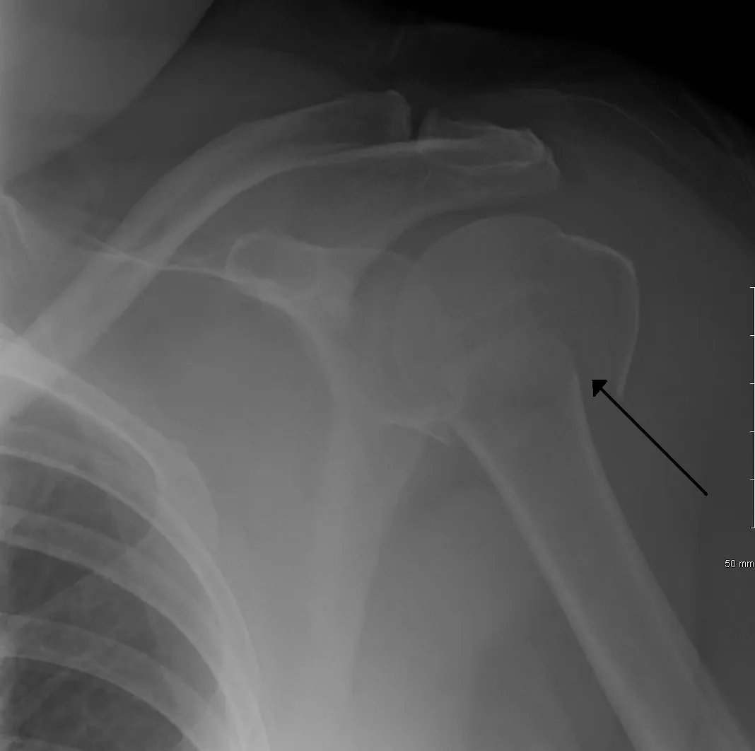
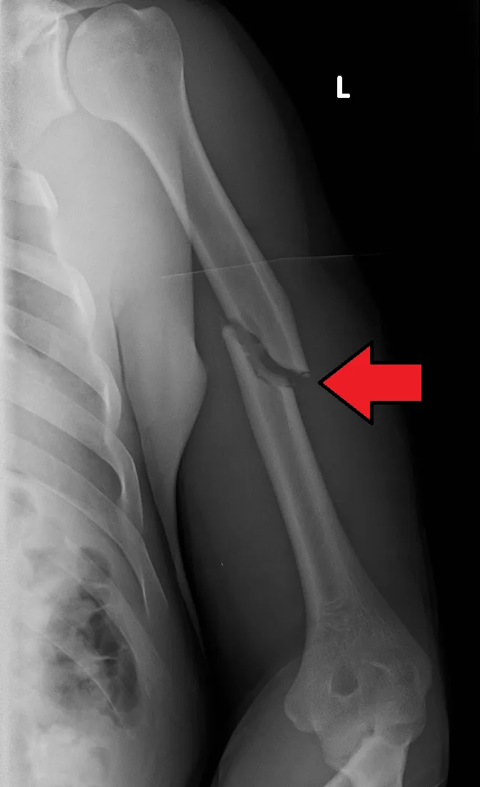
图 3-1 Fig 3-1 真实 X 线:肩关节正位,肱骨外科颈骨折(黑色箭头示骨折线),肱骨头仍位于关节盂内。教学观察点:按头、大结节、小结节、干四部分清点受累节段;但分类不等于手术指征。局限:单一体位;肱骨头血供状态 X 线不可见,缺血风险需结合骨折类型综合判断。独立来源影像,与练习病例无关。Real radiograph: AP shoulder; surgical-neck fracture of the proximal humerus (black arrow marks the fracture line); the humeral head remains seated in the glenoid. Teaching point: count the involved segments (head, greater/lesser tuberosity, shaft) — yet classification is not an operative indication. Limitation: single projection; humeral-head vascularity is not visible on plain radiographs. Independent source image, separate from the practice vignette.图片:James Heilman, MD(Wikimedia Commons),CC BY-SA 3.0(原文件页 · 许可)Image: James Heilman, MD (Wikimedia Commons), CC BY-SA 3.0 (file page · licence)图 3-2 Fig 3-2 真实 X 线:左肱骨正位,骨干中段骨折伴移位(红色箭头为上传者标注)。教学观察点:肱骨干骨折必须在初评及每次操作前后记录桡神经功能(垂腕、虎口区感觉)。局限:神经本身 X 线不可见,神经功能靠查体评估。独立来源影像,与练习病例无关。Real radiograph: AP left humerus; midshaft fracture with displacement (red arrow added by the uploader). Teaching point: document radial nerve function (wrist drop, dorsal first-web sensation) at initial assessment and before/after each manipulation. Limitation: the nerve itself is invisible on radiographs — assess by examination. Independent source image, separate from the practice vignette.图片:James Heilman, MD(Wikimedia Commons),CC BY-SA 4.0(原文件页 · 许可)Image: James Heilman, MD (Wikimedia Commons), CC BY-SA 4.0 (file page · licence)
3.2 初评和红旗3.2 Initial Assessment & Red Flags
视诊触诊:畸形、肿胀、压痛部位定位骨折节段;检查皮肤完整性。Inspection and palpation: deformity, swelling and point tenderness localise the segment; check skin integrity.
三个部位各有必须查的神经:近端—腋神经;骨干—桡神经;远端—尺神经(兼查正中/桡神经)。Each region has its must-check nerve: proximal — axillary; shaft — radial; distal — ulnar (plus median/radial).
血管:触桡动脉搏动;高能量损伤警惕血管损伤。Vascular: palpate the radial pulse; remain vigilant in high-energy injuries.
红旗Red flags
垂腕 / 虎口区麻木:桡神经损伤(骨干骨折最常见)。Wrist drop / dorsal hand numbness: radial nerve injury (most common with shaft fractures).
骨干:按骨折线形态(横形、斜形、螺旋形、粉碎)与部位(上/中/下 1/3)描述。Shaft: described by fracture-line pattern (transverse, oblique, spiral, comminuted) and location (proximal/middle/distal third).
远端:按是否累及关节面、柱的完整性描述(部分关节内 / 完全关节内)。Distal: described by articular involvement and column integrity (partial- vs complete-articular).
以上分类用于沟通与记录;是否手术取决于移位程度、稳定性、患者年龄功能需求与骨质量。These classifications aid communication and documentation; operative decisions depend on displacement, stability, the patient's age and functional demands, and bone quality.
3.4 AO 原则如何适用3.4 How AO Principles Apply
近端:血供保护优先——肱骨头缺血坏死是主要顾虑,粉碎严重时固定与血供之间需权衡。Proximal: blood-supply preservation first — humeral head avascular necrosis is the main concern; weigh fixation against perfusion in severe comminution.
骨干:根据骨折模式、软组织条件、伴随损伤与患者功能需求,在功能支具保守与手术之间选择;两者都是合理选项,关键是与适应证匹配。Shaft: choose between functional bracing and surgery based on fracture pattern, soft-tissue condition, associated injuries and the patient's functional demands; both are legitimate options — matching the indication is what matters.
远端(成人关节内):解剖复位 + 双柱稳定固定 + 早期活动,三者缺一不可。Distal (adult intra-articular): anatomic reduction + bicolumnar stable fixation + early motion — all three are essential.
3.5 治疗选择与限制3.5 Treatment Options & Limits
近端Proximal多数可保守,手术看移位与患者Mostly non-operative; surgery depends on displacement & patient
无移位 / 轻度移位:悬吊制动后渐进康复,多数愈合良好。Non-displaced / minimally displaced: sling then progressive rehabilitation; most heal well.
移位明显的两部分、三部分骨折:年轻、骨质量好 → 多考虑切开复位内固定。Displaced two- or three-part fractures: young patients with good bone → usually open reduction and internal fixation.
高龄、粉碎、骨质量差的四部分骨折:固定困难时可考虑关节置换,个体化决策。Elderly patients with comminuted four-part fractures and poor bone: arthroplasty may be considered when fixation is unfeasible — individualised.
骨干Shaft按骨折与患者情况选择支具或手术,警惕桡神经Brace or surgery by fracture and patient factors; watch the radial nerve
功能支具 / 悬吊 + 早期肩肘活动:适用于骨折模式稳定、软组织条件好、无伴随损伤需手术者。Functional bracing / sling + early shoulder-elbow motion: for stable fracture patterns, good soft-tissue conditions and no associated injuries requiring surgery.
原发性桡神经损伤:多为牵拉伤,观察随访为主,记录基线功能。Primary radial nerve injury: usually a traction injury — observe with follow-up; document baseline function.
手术指征(示例非穷尽):开放骨折、血管损伤需修复、浮肘、多发伤需早期活动、复位后无法维持可接受对线。Operative indications (not exhaustive): open fracture, vascular injury needing repair, floating elbow, polytrauma needing early mobilisation, unacceptable alignment that cannot be maintained.
远端Distal成人关节内多需手术Adult intra-articular often operative
关节外 / 轻度移位:可保守,但需保证早期活动防僵硬。Extra-articular / minimally displaced: may be non-operative, but ensure early motion to prevent stiffness.
成人关节内移位骨折:解剖复位 + 内外侧柱稳定固定,术后早期活动。Displaced adult intra-articular fractures: anatomic reduction + stable medial-lateral column fixation, then early motion.
术中保护尺神经;粉碎严重的高龄患者可个体化考虑置换选项。Protect the ulnar nerve intra-operatively; in severely comminuted elderly fractures, replacement may be individualised.
展开:限制与注意事项Expand: limits and cautions
本课程不给出具体成角/短缩数值标准;“可接受对线”按 AO Surgery Reference 与本院流程判断。This course gives no numeric angulation/shortening cut-offs; "acceptable alignment" follows AO Surgery Reference and local protocols.
本页提供有来源的参考数字与关键步骤:实习识别与报告;规培在上级监督下执行;主治指挥与资源调度。剂量与阈值;以本院规范为准。;开放性骨折用药遵循本院流程。No drug doses or antibiotic regimens are given here; follow local protocols for open fractures.
三个部位不共用同一治疗方案,决策时回到各部位的解剖与功能目标。The three regions do not share one protocol; return to each region's anatomy and functional goals when deciding.
3.6 并发症 / 随访3.6 Complications / Follow-up
近端:肱骨头缺血坏死、骨折不愈合/畸形愈合、肩关节僵硬。Proximal: humeral head avascular necrosis, nonunion/malunion, shoulder stiffness.
随访:X 线愈合进展、神经功能恢复、关节活动度;康复贯穿全程。Follow-up: radiographic healing, nerve recovery, range of motion; rehabilitation throughout.
3.7 病例自测3.7 Case Self-Test
病例 3-1(分段揭晓)Case 3-1 (step-by-step reveal)
注:本病例为合成教学病例,非真实患者。Note: synthetic teaching case, not a real patient.
60 岁男性,车祸致左上臂剧痛、畸形。逐步揭晓。A 60-year-old man has severe left upper-arm pain and deformity after a road traffic crash. Reveal step by step.
步骤 1 · 查体Step 1 · Exam 左上臂中段肿胀畸形,皮肤完整。垂腕,虎口区感觉减退,桡动脉搏动可及。Swelling and deformity of the left mid-upper arm; skin intact. Wrist drop with reduced dorsal-hand sensation; radial pulse palpable.
步骤 2 · 影像Step 2 · Imaging X 线:肱骨中段横形骨折,轻度成角,无明显粉碎。诊断:肱骨干骨折合并桡神经损伤(原发性)。Radiographs: transverse midshaft humeral fracture, mild angulation, no major comminution. Diagnosis: humeral shaft fracture with primary radial nerve injury.
步骤 3 · 决策Step 3 · Decision 骨折本身可保守(功能支具);桡神经多为牵拉伤,先观察。记录基线神经功能,支具固定后早期肩肘活动,定期复查神经恢复与骨折愈合。The fracture itself suits non-operative care (functional bracing); the radial nerve injury is likely tractional — observe first. Document baseline nerve function, brace, begin early shoulder-elbow motion, and follow nerve recovery and union.
步骤 4 · 何时升级Step 4 · When to escalate 若为开放骨折、血管损伤、Holstein-Lewis 型嵌顿可疑或长期不愈合,则转手术。本例先保守+严密随访。Escalate to surgery for open fracture, vascular injury, suspected Holstein-Lewis entrapment or persistent nonunion. This case: non-operative first with close follow-up.
Q1. 肱骨干骨折合并垂腕,最合适的初始处理是?Q1. What is the most appropriate initial management of a humeral shaft fracture with wrist drop?
答案:CAnswer: C 原发性桡神经损伤多为牵拉伤,可自行恢复;关键是记录基线以便随访对比。本例为横形、中段、轻度成角、无明显粉碎,适合功能支具;若为开放骨折、血管损伤或 Holstein-Lewis 嵌顿可疑,则按手术指征处理——选择取决于骨折模式与伴随情况,而非"一律保守"。Primary radial nerve injuries are usually tractional and recover; the key is documenting baseline function for comparison. This case — transverse, midshaft, mild angulation, no major comminution — suits functional bracing; open fracture, vascular injury or suspected Holstein-Lewis entrapment would follow operative indications instead. The choice depends on fracture pattern and associated conditions, not a blanket "always non-operative".
Q2. 关于肱骨三个部位治疗逻辑,正确的是?Q2. Which statement about treatment logic across the three humeral regions is correct?
答案:BAnswer: B 分类描述损伤,不等于手术指征;三个部位解剖与功能目标不同,治疗方案不能套用。Classification describes injury, not surgical indication; the three regions differ in anatomy and functional goals, so protocols cannot be copy-pasted.
AO Surgery Reference — 肱骨远端解剖概念 远端双柱与关节面的官方解剖讲解Official anatomic teaching on the distal humeral columns and articular surface
PubMed — Hettrich 2010 肱骨头血供 肱骨头由旋肱前/后动脉及吻合网共同供血的尸体定量研究Cadaveric quantitative study: humeral head supplied jointly by anterior/posterior circumflex arteries and anastomoses
注:依 AO FAQ,AO 图例仅允许非商业演示使用;在网站转载需申请许可。本课程仅外链官方原页面,未复制、未热链任何 AO 图片。Note: per the AO FAQ, AO illustrations may be used in non-commercial presentations only; republication on a website needs permission. This course links to the official pages and copies or hotlinks no AO images.
本单元来源与媒体许可Sources & media licence — Unit 3
注:“来源(已核验)”仅指该网页于访问日期可正常打开、标题与定位描述相符;不构成对本课程每条医学表述的逐条医学审核。临床决策请结合最新指南与本院流程。Note: "Source (verified)" means only that the page was accessible on the access date and that its title and scope description match; it does not constitute item-by-item medical review of every statement in this course. Base clinical decisions on current guidelines and local protocols.
来源 2(已核验 · 骨干)Source 2 (verified · shaft) 12A1 Simple fracture, spiral — definition 定位:肱骨干螺旋形骨折定义页(AO Surgery Reference,成人创伤模块);按近 / 中 / 远 1/3 分区,明确远 1/3 桡神经风险更高;支撑骨干分类与“桡神经红旗”。Scope: humeral shaft spiral fracture definition page (AO Surgery Reference, adult trauma); zones by proximal/middle/distal third, noting higher radial-nerve risk in the distal third; supports shaft classification and the radial-nerve red flag. 访问日期:2026-10-04Accessed: 2026-10-04
来源 3(已核验 · 远端)Source 3 (verified · distal) Anatomical concepts (distal humerus) 定位:肱骨远端解剖概念页(AO Surgery Reference,成人创伤模块);阐述内外侧柱三角稳定结构等解剖/生物力学概念,支撑“解剖与机制”。注意此为解剖页,非某型骨折的治疗页。Scope: distal humerus anatomical concepts page (AO Surgery Reference, adult trauma); covers the triangular columnar stability construct and related biomechanics; supports "Anatomy & Mechanism". Note: an anatomy page, not a fracture-subtype treatment page. 访问日期:2026-10-04Accessed: 2026-10-04
来源 4(已核验 · 解剖研究)Source 4 (verified · anatomic study) Quantitative assessment of the vascularity of the proximal part of the humerus 定位:Hettrich 等,J Bone Joint Surg Am 2010;24 具尸体肩的定量研究:后旋肱动脉对肱骨头的灌注贡献大于既往认识,结论建议手术入路与固定时保护后旋肱动脉。课程仅引用其“后旋肱动脉贡献大于既往认识、应保护软组织附着”的结论,不把该研究的比例数字外推为所有患者的固定数值。Scope: Hettrich et al., J Bone Joint Surg Am 2010; quantitative study in 24 cadaver shoulders: the posterior humeral circumflex artery contributes more perfusion to the humeral head than previously described; the authors advise protecting it during approach and fixation. This course cites only the "posterior vessel matters more than previously thought; protect soft-tissue attachments" conclusion, without extrapolating the study's proportions to all patients. 访问日期:2026-10-04Accessed: 2026-10-04
诚实标注:肱骨远端各骨折亚型的具体术式 treatment 页面未逐页打开核验,本单元未引用任何具体术式页;治疗选择章节仅给原则性分层。Honesty note: subtype-specific operative treatment pages for distal humerus fractures were not individually opened and verified; this unit cites no specific operative technique pages, and the treatment section stays at the level of principles.
本单元图片(图 3-1、图 3-2):真实 X 线/CT,逐图核验许可;作者、原标题、原 URL、许可与原 caption 见 assets/media_sources.csv,各图注含原文件页与许可链接。各图为不同患者的独立图片,非同一病例序列。Unit images (Figs 3-1, 3-2): real radiographs/CT, licences verified per image; author, original title, source URL, licence and original caption are listed in assets/media_sources.csv, and each caption links its file page and licence. Images are from different patients, not a case sequence.
本单元铁律Unit rule 每一例前臂骨折,都要看肘、看腕。Monteggia 漏看肘、Galeazzi 漏看腕,是前臂创伤最经典的漏诊。For every forearm fracture, examine the elbow and the wrist. Missing the elbow in Monteggia and the wrist in Galeazzi are the classic missed diagnoses of forearm trauma.
4.1 解剖与机制4.1 Anatomy & Mechanism
尺桡骨通过上尺桡关节、骨间膜、下尺桡关节(DRUJ)连成一个旋转整体,前臂旋转功能依赖两骨的相对长度与对线。Radius and ulna form one rotational unit via the proximal radioulnar joint, interosseous membrane and DRUJ; forearm rotation depends on their relative length and alignment.
双骨折(尺桡骨干双骨折)多为高能量直接暴力。Both-bone forearm fractures usually result from high-energy direct trauma.
Monteggia:尺骨近端骨折 + 桡骨头脱位(看肘)。Monteggia: proximal ulna fracture + radial head dislocation (check the elbow).
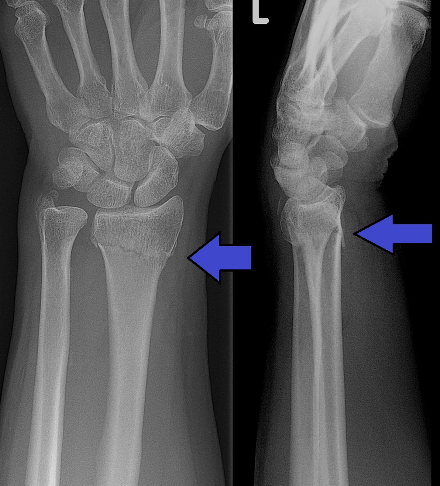
桡骨远端骨折:跌倒手撑地最常见;关注关节面、DRUJ 与腕管。Distal radius fracture: most commonly a fall onto the outstretched hand; assess the articular surface, DRUJ and carpal tunnel.
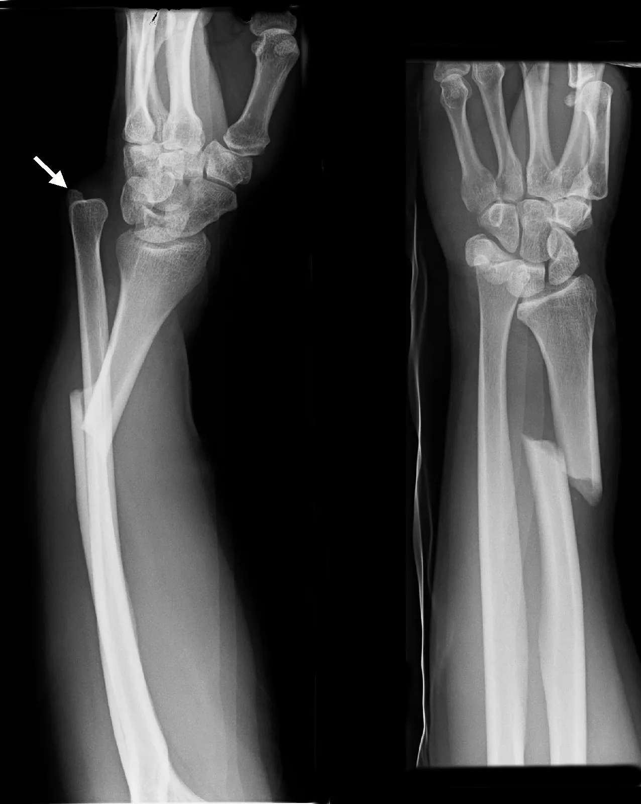
图 4-1 Fig 4-1 真实 X 线(正侧位):桡骨干远端短斜行骨折,合并远端尺桡关节完全脱位(白色箭头示脱位的尺骨头)——Galeazzi 损伤。教学观察点:桡骨干骨折必须检查腕关节(DRUJ),尺骨干骨折必须检查肘关节。局限:单病例;DRUJ 稳定性需结合查体与专科评估。独立来源影像,与练习病例无关。Real radiographs (AP and lateral): short oblique fracture of the distal radial shaft with complete dislocation of the distal radioulnar joint (white arrow marks the dislocated ulnar head) — a Galeazzi injury. Teaching point: always examine the wrist (DRUJ) with a radial shaft fracture, and the elbow with an ulnar shaft fracture. Limitation: single case; DRUJ stability needs clinical assessment. Independent source image, separate from the practice vignette.图片:Th. Zimmermann(Wikimedia Commons),CC BY-SA 3.0 DE(原文件页 · 许可)Image: Th. Zimmermann (Wikimedia Commons), CC BY-SA 3.0 DE (file page · licence)图 4-2 Fig 4-2 真实 X 线(正侧位):左腕 Colles 骨折——桡骨远端骨折伴背侧移位(蓝色箭头为上传者标注),合并尺骨茎突骨折。教学观察点:评估关节面是否受累、DRUJ 是否稳定、正中神经与骨筋膜室红旗。局限:DRUJ 受累本身不等于手术指征,需结合稳定性综合判断。独立来源影像,与练习病例无关。Real radiographs (AP and lateral): Colles fracture of the left wrist — dorsally displaced distal radius fracture (blue arrows added by the uploader) with an ulnar styloid fracture. Teaching point: assess articular involvement, DRUJ stability, and median-nerve / compartment red flags. Limitation: DRUJ involvement alone is not an operative indication — judge stability comprehensively. Independent source image, separate from the practice vignette.图片:Lucien Monfils;箭头标注:Doc James(Wikimedia Commons),CC BY-SA 3.0(原文件页 · 许可)Image: Lucien Monfils; arrows: Doc James (Wikimedia Commons), CC BY-SA 3.0 (file page · licence)展开:为什么前臂双骨折多需手术?Expand: why do both-bone forearm fractures often need surgery?
前臂旋转功能对两骨长度与成角极敏感,闭合复位难以同时维持两骨的解剖对线。Forearm rotation is highly sensitive to the length and angulation of both bones; closed reduction struggles to hold anatomic alignment of both.
畸形愈合 → 旋转受限,这是前臂骨折区别于肱骨干骨折的关键。Malunion → loss of rotation. This is what sets forearm fractures apart from humeral shaft fractures.
4.2 初评和红旗4.2 Initial Assessment & Red Flags
查骨折局部:畸形、肿胀、压痛、皮肤完整性。Examine the fracture site: deformity, swelling, tenderness, skin integrity.
查肘:桡骨头有无脱位(Monteggia)。查腕:DRUJ 有无脱位、腕部压痛(Galeazzi)。Check the elbow: radial head dislocation (Monteggia). Check the wrist: DRUJ dislocation, wrist tenderness (Galeazzi).
急性腕管综合征:桡骨远端骨折后正中神经分布区进行性麻木、疼痛加剧。Acute carpal tunnel syndrome: progressive median-nerve numbness and worsening pain after distal radius fracture.
骨筋膜室综合征:前臂张力性肿胀、被动牵拉痛。Compartment syndrome: tense forearm swelling, pain on passive stretch.
开放性骨折 / 皮肤破损。Open fracture / skin breach.
单骨骨折 X 线:必须追看肘与腕,否则 Monteggia / Galeazzi 漏诊。Single-bone fracture on X-ray: must review elbow and wrist, or Monteggia/Galeazzi will be missed.
4.3 影像 / 分类4.3 Imaging / Classification
常规:前臂正侧位 X 线,必须包含肘关节与腕关节。Routine: AP and lateral forearm radiographs — must include the elbow and the wrist.
桡骨远端:加腕关节正侧位,评估关节面台阶、DRUJ 对合。Distal radius: add wrist AP/lateral views to assess articular step and DRUJ congruity.
复杂关节内骨折:CT 明确关节面粉碎与移位。Complex intra-articular fractures: CT to define articular comminution and displacement.
展开:分类概念Expand: classification concepts
骨干:单骨 vs 双骨;骨折线形态与部位;是否合并关节脱位(Monteggia / Galeazzi 为特殊类型)。Shaft: single- vs both-bone; fracture-line pattern and location; associated joint dislocation (Monteggia/Galeazzi as special types).
远端:关节外 vs 部分关节内 vs 完全关节内;DRUJ 是否受累。Distal: extra-articular vs partial-articular vs complete-articular; DRUJ involvement or not.
分类用于描述,不等于手术指征。Classification describes; it is not an operative indication.
4.4 AO 原则如何适用4.4 How AO Principles Apply
解剖复位在这里有功能含义:恢复两骨长度、对线与旋转对合,不只是“接上”。Anatomic reduction here has functional meaning: restoring length, alignment and rotational congruity of both bones — not just "joining" them.
稳定固定:为早期功能锻炼创造条件;但若合并 DRUJ 损伤(如 Galeazzi),韧带愈合需优先保护,旋转锻炼须待稳定性确认后进行,不可过早自由旋转。Stable fixation: creates the conditions for early functional exercise; but with DRUJ injury (e.g. Galeazzi), ligament healing takes priority — rotational exercises only after stability is confirmed, never early free rotation.
保护血供:前臂软组织包被少,手术入路注意保护皮神经与肌腱。Preserve blood supply: thin soft-tissue envelope — protect cutaneous nerves and tendons in surgical approaches.
Monteggia / Galeazzi:复位的核心是恢复脱位关节的对合,骨折固定为关节稳定服务。Monteggia/Galeazzi: the core of reduction is restoring the dislocated joint; fracture fixation serves joint stability.
4.5 治疗选择与限制4.5 Treatment Options & Limits
骨干双骨折Both-bone shaft成人多需手术恢复旋转功能Adults often need surgery to restore rotation
成人尺桡骨干双骨折:多需切开复位内固定,恢复两骨解剖对线。Adult both-bone diaphyseal fractures: usually open reduction and internal fixation to restore anatomic alignment of both bones.
术后在骨折稳定固定、且 DRUJ 无损伤的前提下,早期开始前臂旋转锻炼。After surgery, provided fixation is stable and the DRUJ is intact, begin forearm rotation exercises early.
Monteggia / GaleazziMonteggia / Galeazzi先认出,再按关节处理Recognise first, then treat the joint
Monteggia:复位固定尺骨骨折,恢复桡骨头对合;检查桡骨头稳定性与骨间后神经。Monteggia: reduce and fix the ulna fracture, restore radial head congruity; check radial head stability and the posterior interosseous nerve.
Galeazzi:复位固定桡骨骨折,评估 DRUJ 稳定性;DRUJ 不稳定需同期处理。术后将前臂置于 DRUJ 最稳定的旋转体位制动以保护韧带愈合,旋转锻炼待稳定性确认后再开始。Galeazzi: reduce and fix the radius fracture, assess DRUJ stability; unstable DRUJ needs concurrent management. Post-operatively, splint the forearm in the rotational position where the DRUJ is most stable to protect ligament healing; rotational exercises only after stability is confirmed.
两者都不是“单纯前臂骨折”,漏诊即治疗错误。Neither is "just a forearm fracture" — missing the diagnosis means wrong treatment.
桡骨远端Distal radius按关节受累与稳定性分层Stratify by articular involvement & stability
关节外、稳定:闭合复位 + 石膏/支具,多可保守。Extra-articular and stable: closed reduction + cast/brace; often non-operative.
关节内明显移位或整体不稳定:多需手术恢复关节面。Markedly displaced intra-articular fractures or overall instability: usually operative to restore the articular surface.
DRUJ 受累本身不等于手术指征:仅受累而稳定者可保守并随访;持续不稳定或移位者,结合年龄、功能需求、关节面情况与整体稳定性综合决定。DRUJ involvement alone is not a surgical indication: stable involvement may be managed non-operatively with follow-up; persistent instability or displacement calls for an individualised decision integrating age, functional demands, articular status and overall stability.
本课程不给出具体成角数值标准与内固定型号选择,遵循 AO Surgery Reference 与本院流程。No numeric angulation cut-offs or implant choices are given; follow AO Surgery Reference and local protocols.
急性腕管综合征是急症:进行性正中神经症状需紧急处理,不要等待观察。Acute carpal tunnel syndrome is an emergency: progressive median-nerve symptoms need urgent management, not watchful waiting.
4.6 并发症 / 随访4.6 Complications / Follow-up
旋转功能受限:前臂骨折最特征的并发症,康复重点。Loss of rotation: the signature complication of forearm fractures; the focus of rehabilitation.
骨不连 / 畸形愈合:尤其双骨折对线不良者。Nonunion / malunion: especially with poor alignment in both-bone fractures.
注:本病例为合成教学病例,非真实患者。Note: synthetic teaching case, not a real patient.
30 岁男性,跌倒后左前臂剧痛、畸形。逐步揭晓。A 30-year-old man has severe left forearm pain and deformity after a fall. Reveal step by step.
步骤 1 · 查体Step 1 · Exam 前臂中段畸形,皮肤完整,桡动脉搏动可及。按铁律查肘查腕:肘部压痛不明显,腕部尺侧压痛明显、DRUJ 区饱满。Mid-forearm deformity; skin intact; radial pulse palpable. Per the unit rule, check elbow and wrist: elbow non-tender, but marked ulnar-sided wrist tenderness with a prominent DRUJ.
步骤 2 · 影像Step 2 · Imaging 前臂正侧位(含肘腕):桡骨干中段骨折、成角移位;腕关节 DRUJ 间隙增宽、尺骨头向背侧半脱位。诊断:Galeazzi 损伤。Forearm AP/lateral including elbow and wrist: midshaft radial fracture with angulation; widened DRUJ with dorsal subluxation of the ulnar head. Diagnosis: Galeazzi injury.
步骤 3 · 决策Step 3 · Decision 这不是单纯桡骨骨折。手术复位固定桡骨,术中评估 DRUJ 稳定性;若 DRUJ 不稳定,同期稳定 DRUJ。术后将前臂置于 DRUJ 最稳定的旋转体位制动(控制旋转,促进韧带愈合),旋转锻炼待稳定性确认后再开始;若 DRUJ 明显不稳定需克氏针临时固定,则制动期间禁止前臂旋转。Not just a radial fracture. Operative reduction and fixation of the radius with intra-operative DRUJ stability assessment; stabilise the DRUJ concurrently if unstable. Post-operatively, splint the forearm in the rotational position where the DRUJ is most stable (controlling rotation to allow ligament healing); begin rotational exercises only after stability is confirmed. If gross DRUJ instability requires temporary K-wire transfixation, forearm rotation is prohibited during immobilisation.
步骤 4 · 教训Step 4 · Lesson 若只看前臂正侧位中央段、X 线未包含腕关节,DRUJ 脱位将被漏诊,单纯固定桡骨会导致腕关节慢性不稳定。Had the radiograph not included the wrist, the DRUJ dislocation would have been missed, and fixing only the radius would leave chronic wrist instability.
Q1. 前臂 X 线显示单纯尺骨干骨折,接下来的关键动作是?Q1. Forearm radiographs show an isolated ulnar shaft fracture. What is the key next step?
答案:BAnswer: B 尺骨骨折 + 桡骨头脱位 = Monteggia。单骨骨折必须看相邻关节,这是前臂创伤的铁律。Ulna fracture + radial head dislocation = Monteggia. Single-bone fractures demand assessment of the adjacent joint — the iron rule of forearm trauma.
Q2. 桡骨远端骨折复位固定后,患者诉拇指、示指进行性麻木、胀痛加重,应首先考虑?Q2. After reduction and fixation of a distal radius fracture, the patient reports progressive thumb/index numbness and worsening swelling pain. First consideration?
答案:DAnswer: D 进行性正中神经分布区麻木 + 疼痛加重是红旗,需立即评估腕管压力与前臂骨筋膜室,必要时紧急减压。等待观察可致不可逆损伤。Progressive median-nerve numbness with worsening pain is a red flag — urgently assess carpal tunnel pressure and forearm compartments; decompress emergently if needed. Waiting risks irreversible damage.
注:依 AO FAQ,AO 图例仅允许非商业演示使用;在网站转载需申请许可。本课程仅外链官方原页面,未复制、未热链任何 AO 图片。Note: per the AO FAQ, AO illustrations may be used in non-commercial presentations only; republication on a website needs permission. This course links to the official pages and copies or hotlinks no AO images.
本单元来源与媒体许可Sources & media licence — Unit 4
注:“来源(已核验)”仅指该网页于访问日期可正常打开、标题与定位描述相符;不构成对本课程每条医学表述的逐条医学审核。临床决策请结合最新指南与本院流程。Note: "Source (verified)" means only that the page was accessible on the access date and that its title and scope description match; it does not constitute item-by-item medical review of every statement in this course. Base clinical decisions on current guidelines and local protocols.
来源 1(已核验 · Monteggia)Source 1 (verified · Monteggia) Simple fracture of the ulna, with dislocation of proximal radioulnar joint (Monteggia) 定位:Monteggia 损伤治疗模块入口页(AO Surgery Reference,成人创伤模块,含诊断 → 适应证 → 治疗导航);支撑“相邻关节受累”与 Monteggia 章节。子术式页未逐页点开。Scope: Monteggia injury treatment-module entry page (AO Surgery Reference, adult trauma, with diagnosis → indications → treatment navigation); supports the adjacent-joint and Monteggia sections. Sub-technique pages were not individually opened. 访问日期:2026-10-04Accessed: 2026-10-04
来源 2(已核验 · Galeazzi)Source 2 (verified · Galeazzi) Simple fracture of the radius, with dislocation of distal radioulnar joint (Galeazzi) 定位:Galeazzi 损伤治疗模块入口页(AO Surgery Reference,成人创伤模块);支撑 Galeazzi 章节与 DRUJ 稳定性评估。子术式页未逐页点开。Scope: Galeazzi injury treatment-module entry page (AO Surgery Reference, adult trauma); supports the Galeazzi section and DRUJ stability assessment. Sub-technique pages were not individually opened. 访问日期:2026-10-04Accessed: 2026-10-04
来源 3(已核验 · 桡骨远端)Source 3 (verified · distal radius) ORIF - Palmar plate (partial articular fracture of the radius, involving the volar rim) 定位:桡骨远端掌侧缘部分关节内骨折的掌侧接骨板 ORIF 页(AO Surgery Reference,成人创伤模块);含复位原则、DRUJ 稳定性测试方法、术后随访;支撑治疗选择与随访章节。Scope: palmar-plate ORIF page for partial-articular volar-rim distal radius fractures (AO Surgery Reference, adult trauma); covers reduction principles, DRUJ stability testing and post-operative follow-up; supports the treatment and follow-up sections. 访问日期:2026-10-04Accessed: 2026-10-04
来源 4(已核验 · Galeazzi DRUJ 处理)Source 4 (verified · Galeazzi DRUJ management) DRUJ stabilization (Galeazzi fracture-dislocation) 定位:Galeazzi 损伤 DRUJ 稳定性处理基本技术页(AO Surgery Reference,成人创伤模块);明确稳定性确认后建议以控制旋转的可拆卸支具制动 DRUJ 约 3 周以促进韧带愈合,术后制动取 DRUJ 最稳定的旋转体位,明显不稳定者克氏针临时固定 3–4 周并禁止旋转;支撑本单元 v2 康复修正(保护 DRUJ 韧带愈合、依稳定性限制旋转)。Scope: basic-technique page on DRUJ stabilisation in Galeazzi fracture-dislocation (AO Surgery Reference, adult trauma); once stability is assured, immobilise the DRUJ ~3 weeks in a rotation-controlling removable splint to allow ligament healing, splint in the most stable rotational position, and for gross instability use temporary K-wire transfixation for 3–4 weeks with rotation prohibited; supports this unit's v2 rehabilitation correction (protect DRUJ ligament healing; limit rotation by stability). 访问日期:2026-10-04Accessed: 2026-10-04
本单元图片(图 4-1、图 4-2):真实 X 线/CT,逐图核验许可;作者、原标题、原 URL、许可与原 caption 见 assets/media_sources.csv,各图注含原文件页与许可链接。各图为不同患者的独立图片,非同一病例序列。Unit images (Figs 4-1, 4-2): real radiographs/CT, licences verified per image; author, original title, source URL, licence and original caption are listed in assets/media_sources.csv, and each caption links its file page and licence. Images are from different patients, not a case sequence.
临床判断与关键步骤Clinical decisions and key steps
具体方案;以本院为准;每项数字同时核对人群、阶段和来源。Follow institutional protocols; check population, stage and source with every number.
开放骨折:抗生素、清创与覆盖Open fracture: antibiotics, debridement and coverage
抗生素依据并列Antibiotic sources side by side
尽早静脉抗生素,尽量伤后1 h内。国内2019 Gustilo I–II以一代头孢(原文头孢唑啉)为基础;III加氨基糖苷,粪便/厌氧污染加青霉素或甲硝唑;过敏/肾功能及本地耐药谱决定替代。以本院规范为准。Early IV antibiotics, ideally within one hour. Chinese 2019 GustiloI–II first-generation cephalosporin(cefazolin), III adds aminoglycoside, fecal/anaerobic contamination adds penicillin/metronidazole; allergy/renal function/local resistance guide alternatives. Follow institutional policy.
国内2019持续至闭合后48 h;不能当所有现代方案统一疗程,按本院抗菌管理选择。盐水湿敷+封闭层与授权照片;BOAST/NICE不做急诊小冲洗,国内旧文急诊清洗说法另需本院定标。以本院规范为准。Chinese 2019 continues until48 hours after closure, not a universal modern duration; use local stewardship. Saline-moist/occlusive dressing and authorized photo; BOAST/NICE no ED mini-washout differs from older Chinese cleaning text. Follow institutional policy.
清创与重建Debridement and reconstruction
重污染/血管受损立即;其他高能12 h内、低能24 h内清创;确定覆盖尽量72 h内,内固定需与覆盖配合。破伤风与多区域复评同步。以本院规范为准。Immediate debridement for severe contamination/vascular compromise; other high-energy within 12 hours/low-energy within 24; definitive coverage ideally within 72, coordinated with fixation. Include tetanus and whole-patient review. Follow institutional policy.
筋膜室:早诊与立即减压Compartment syndrome: early diagnosis and immediate decompression
有脉搏也可缺血A pulse can coexist with ischemia
高风险每小时查体,记录疼痛/被动牵拉痛及镇痛剂量;有症状松解环形层至皮肤,心脏水平,30 min内复评并即时骨科会诊。以本院规范为准。High risk: hourly exam, pain/passive stretch/analgesic dose. Symptoms: release circumferential layers to skin, heart-level limb, reassess within 30 minutes and consult orthopedics immediately. Follow institutional policy.
DBP−间室压<30 mmHg风险增加;绝对压>40 mmHg考虑紧急减压,不能脱离临床。确诊后立即切开;国内2019“6–8 h内”是旧表述,不能作为等待理由。以本院规范为准。DBP minus compartment pressure below 30 increases risk; absolute over 40 prompts urgent decompression assessment in context. Once diagnosed, immediate fasciotomy; older Chinese 2019 wording within 6–8 hours is not permission to delay. Follow institutional policy.
肢体血管伤:没有安全等待时间Limb vascular injury: no safe waiting time
硬征与紧急复流Hard signs and urgent reperfusion
无脉、持续出血、扩张性血肿为硬征;毛细血管再充盈或Doppler不能单独排除。立即骨科/血管/整形协同,必要时临时分流。以本院规范为准。Absent pulse/ongoing bleeding/expanding hematoma are hard signs; CRT/Doppler alone cannot exclude injury. Immediate orthopedic/vascular/plastic coordination, temporary shunt when needed. Follow institutional policy.
BOAST2026:到院1 h内开始血运重建(commenced),不是承诺1 h内已恢复;不因复杂影像延误复流。以本院规范为准。BOAST 2026: commence revascularization within one hour of arrival, not a guarantee of completed flow restoration by then; complex imaging must not delay reperfusion. Follow institutional policy.
本区重点:查体、血供与升级Regional focus: examination, perfusion and escalation
上肢:前后对照查体Upper limb: pre/post comparison
肱骨髁上骨折/肘脱位:记录桡动脉、指端灌注及正中(含骨间前)、桡、尺神经运动与感觉;复位前后逐项对照(BOAST)。Supracondylar fracture/elbow dislocation: document radial pulse, digital perfusion and median (including AIN), radial and ulnar motor/sensory findings before and after reduction (BOAST).
肱骨干骨折:复位前后记录伸腕/伸指及虎口背侧感觉;新桡神经缺损立即升级,不能只凭复位片满意结束(BOAST神经血管原则)。Humeral shaft fracture: record wrist/finger extension and dorsal first-web sensation before/after reduction. Escalate new radial deficits despite satisfactory alignment (BOAST neurovascular principles).
前臂高风险:持续疼痛、被动牵拉痛或感觉变化即复评;松解环形束缚、肢体心脏水平,立即骨科评估(BOAST筋膜室)。Forearm risk: worsening pain, passive-stretch pain or sensory change requires reassessment; release constriction, keep at heart level and obtain urgent orthopedic review (BOAST compartment guidance).
上肢用BBI/API(不是踝指数);<0.9或脉搏不对称,稳定者CTA/血管外科评估;硬征或缺血不等指数/CTA延误救治(WTA/BOAST)。Use upper-limb BBI/API rather than an ankle index; below 0.9 or asymmetric pulses warrants CTA/vascular review if stable. Hard signs or ischemia require immediate care without waiting for indices/imaging (WTA/BOAST).